

Quantitative computed tomography determined regional lung mechanics in normal nonsmokers, normal smokers and metastatic sarcoma subjects
- PMID: 28749945
- PMCID: PMC5531492
- DOI: 10.1371/journal.pone.0179812
Quantitative computed tomography determined regional lung mechanics in normal nonsmokers, normal smokers and metastatic sarcoma subjects
Abstract
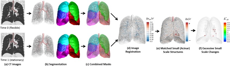
Objectives: Extra-thoracic tumors send out pilot cells that attach to the pulmonary endothelium. We hypothesized that this could alter regional lung mechanics (tissue stiffening or accumulation of fluid and inflammatory cells) through interactions with host cells. We explored this with serial inspiratory computed tomography (CT) and image matching to assess regional changes in lung expansion.
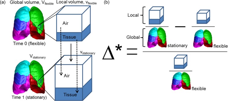
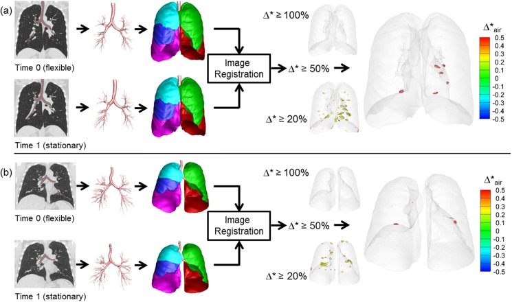
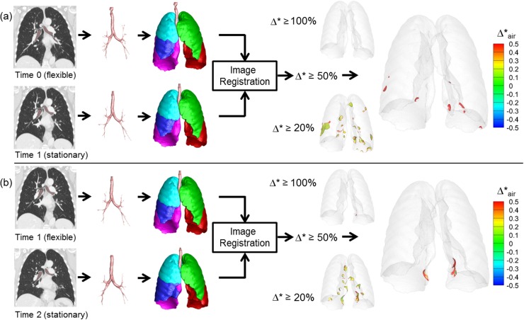
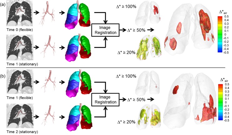
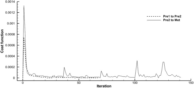
Materials and methods: We retrospectively assessed 44 pairs of two serial CT scans on 21 sarcoma patients: 12 without lung metastases and 9 with lung metastases. For each subject, two or more serial inspiratory clinically-derived CT scans were retrospectively collected. Two research-derived control groups were included: 7 normal nonsmokers and 12 asymptomatic smokers with two inspiratory scans taken the same day or one year apart respectively. We performed image registration for local-to-local matching scans to baseline, and derived local expansion and density changes at an acinar scale. Welch two sample t test was used for comparison between groups. Statistical significance was determined with a p value < 0.05.
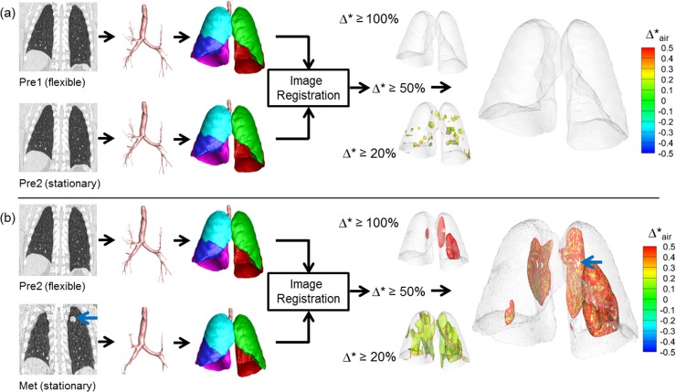
Results: Lung regions of metastatic sarcoma patients (but not the normal control group) demonstrated an increased proportion of normalized lung expansion between the first and second CT. These hyper-expanded regions were associated with, but not limited to, visible metastatic lung lesions. Compared with the normal control group, the percent of increased normalized hyper-expanded lung in sarcoma subjects was significantly increased (p < 0.05). There was also evidence of increased lung "tissue" volume (non-air components) in the hyper-expanded regions of the cancer subjects relative to non-hyper-expanded regions. "Tissue" volume increase was present in the hyper-expanded regions of metastatic and non-metastatic sarcoma subjects. This putatively could represent regional inflammation related to the presence of tumor pilot cell-host related interactions.
Conclusions: This new quantitative CT (QCT) method for linking serial acquired inspiratory CT images may provide a diagnostic and prognostic means to objectively characterize regional responses in the lung following oncological treatment and monitoring for lung metastases.
Conflict of interest statement
Figures
References
-
- Ma J, Niu M, Yang W, Zang L, Xi Y. Role of relaxin-2 in human primary osteosarcoma. Cancer cell international. 2013;13(1):59 doi: 10.1186/1475-2867-13-59 - DOI - PMC - PubMed
-
- Sethi N, Kang Y. Notch signaling: mediator and therapeutic target of bone metastasis. BoneKEy reports. 2012;1:3 doi: 10.1038/bonekey.2012.2 - DOI - PMC - PubMed
-
- Sethi N, Kang Y. Dysregulation of developmental pathways in bone metastasis. Bone. 2011;48(1):16–22. doi: 10.1016/j.bone.2010.07.005 - DOI - PubMed
-
- Sethi N, Kang Y. Unravelling the complexity of metastasis—molecular understanding and targeted therapies. Nature reviews Cancer. 2011;11(10):735–48. doi: 10.1038/nrc3125 - DOI - PMC - PubMed
-
- Sethi N, Kang Y. Notch signalling in cancer progression and bone metastasis. British journal of cancer. 2011;105(12):1805–10. doi: 10.1038/bjc.2011.497 - DOI - PMC - PubMed
MeSH terms
Grants and funding
LinkOut - more resources
Full Text Sources
Other Literature Sources
Medical
