

Increased level and interferon-γ production of circulating natural killer cells in patients with scrub typhus
- PMID: 28750012
- PMCID: PMC5549767
- DOI: 10.1371/journal.pntd.0005815
Increased level and interferon-γ production of circulating natural killer cells in patients with scrub typhus
Abstract
Background: Natural killer (NK) cells are essential immune cells against several pathogens. Not much is known regarding the roll of NK cells in Orientia tsutsugamushi infection. Thus, this study aims to determine the level, function, and clinical relevance of NK cells in patients with scrub typhus.
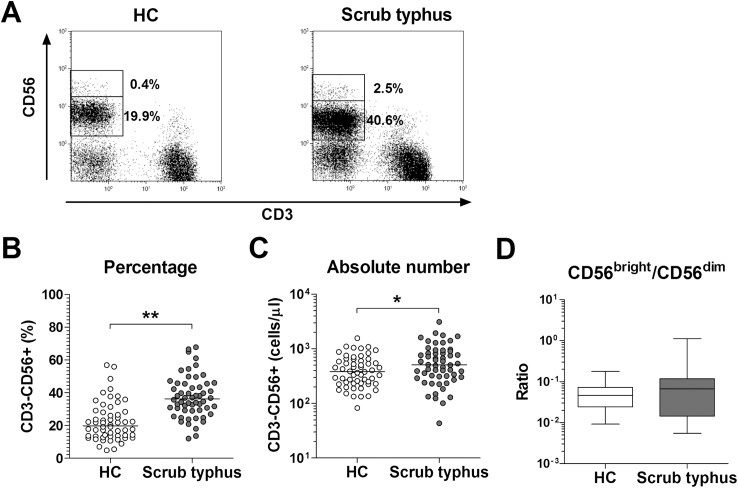
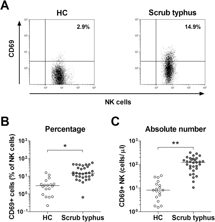
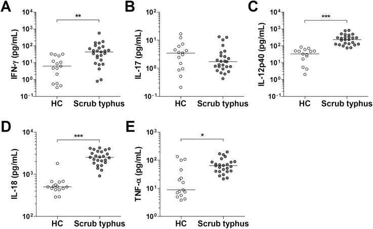
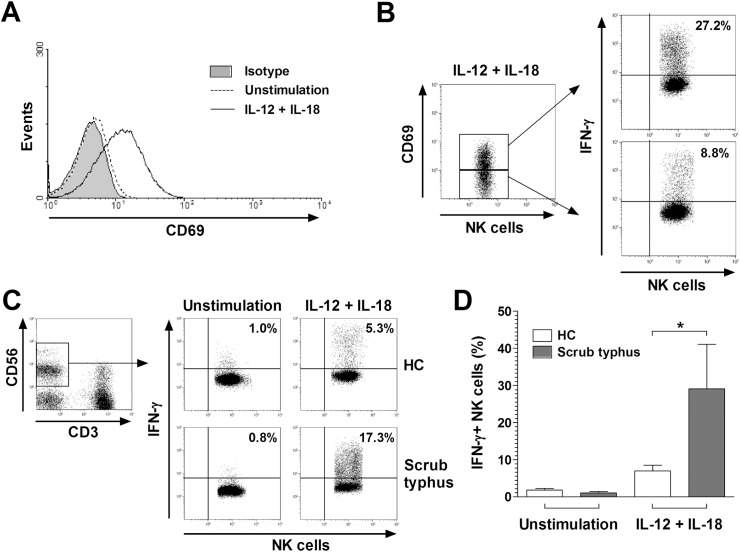
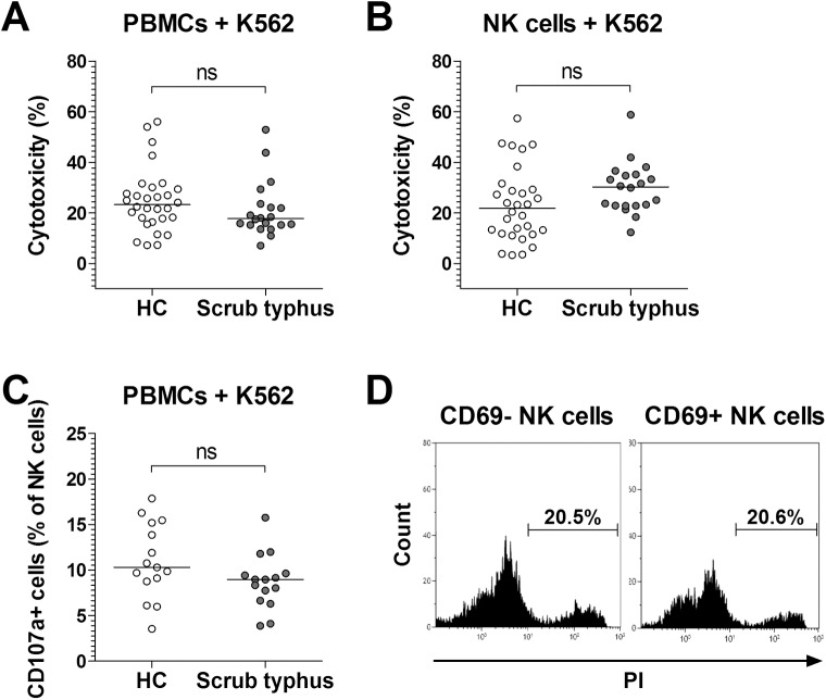
Methodology/principal findings: This study enrolled fifty-six scrub typhus patients and 56 health controls (HCs). The patients were divided into subgroups according to their disease severity. A flow cytometry measured NK cell level and function in peripheral blood. Circulating NK cell levels and CD69 expressions were significantly increased in scrub typhus patients. Increased NK cell levels reflected disease severity. In scrub typhus patients, tests showed their NK cells produced higher amounts of interferon (IFN)-γ after stimulation with interleukin (IL)-12 and IL-18 relative to those of HCs. Meanwhile, between scrub typhus patients and HCs, the cytotoxicity and degranulation of NK cells against K562 were comparable. CD69 expressions were recovered to the normal levels in the remission phase.
Conclusions: This study shows that circulating NK cells are activated and numerically increased, and they produced more IFN-γ in scrub typhus patients.
Conflict of interest statement
The authors have declared that no competing interests exist.
Figures

Similar articles
-
Dysfunction of Circulating Natural Killer T Cells in Patients With Scrub Typhus.J Infect Dis. 2018 Oct 20;218(11):1813-1821. doi: 10.1093/infdis/jiy402. J Infect Dis. 2018. PMID: 29982731
-
Circulating Plasmacytoid and Conventional Dendritic Cells Are Numerically and Functionally Deficient in Patients With Scrub Typhus.Front Immunol. 2021 Jul 1;12:700755. doi: 10.3389/fimmu.2021.700755. eCollection 2021. Front Immunol. 2021. PMID: 34276693 Free PMC article.
-
Phenotype and function of natural killer cells in systemic lupus erythematosus: excess interferon-γ production in patients with active disease.Arthritis Rheum. 2011 Jun;63(6):1698-706. doi: 10.1002/art.30313. Arthritis Rheum. 2011. PMID: 21370226
-
Differential expression of interferon-gamma and interferon-gamma-inducing cytokines in Thai patients with scrub typhus or leptospirosis.Clin Immunol. 2004 Nov;113(2):140-4. doi: 10.1016/j.clim.2004.08.006. Clin Immunol. 2004. PMID: 15451469
-
Dysregulated Th1 Immune and Vascular Responses in Scrub Typhus Pathogenesis.J Immunol. 2018 Feb 15;200(4):1233-1240. doi: 10.4049/jimmunol.1701219. J Immunol. 2018. PMID: 29431689 Free PMC article. Review.
Cited by
-
Distinct Role of TNFR1 and TNFR2 in Protective Immunity Against Orientia tsutsugamushi Infection in Mice.Front Immunol. 2022 Apr 11;13:867924. doi: 10.3389/fimmu.2022.867924. eCollection 2022. Front Immunol. 2022. PMID: 35479068 Free PMC article.
-
Longevity of antibody and T-cell responses against outer membrane antigens of Orientia tsutsugamushi in scrub typhus patients.Emerg Microbes Infect. 2017 Dec 20;6(12):e116. doi: 10.1038/emi.2017.106. Emerg Microbes Infect. 2017. PMID: 29259327 Free PMC article.
-
Natural Killer Cells in Immunotherapy: Are We Nearly There?Cancers (Basel). 2020 Oct 27;12(11):3139. doi: 10.3390/cancers12113139. Cancers (Basel). 2020. PMID: 33120910 Free PMC article. Review.
-
The Obligate Intracellular Bacterium Orientia tsutsugamushi Targets NLRC5 To Modulate the Major Histocompatibility Complex Class I Pathway.Infect Immun. 2019 Feb 21;87(3):e00876-18. doi: 10.1128/IAI.00876-18. Print 2019 Mar. Infect Immun. 2019. PMID: 30559222 Free PMC article.
-
Diagnostic Value of CD4/CD8 in Scrub Typhus.Am J Trop Med Hyg. 2021 Dec 13;106(3):792-797. doi: 10.4269/ajtmh.21-0531. Am J Trop Med Hyg. 2021. PMID: 34902835 Free PMC article.
References
-
- Jensenius M, Fournier PE, Raoult D (2004) Rickettsioses and the international traveler. Clin Infect Dis 39: 1493–1499. doi: 10.1086/425365 - DOI - PubMed
-
- Taylor AJ, Paris DH, Newton PN (2015) A Systematic Review of Mortality from Untreated Scrub Typhus (Orientia tsutsugamushi). PLoS Negl Trop Dis 9: e0003971 doi: 10.1371/journal.pntd.0003971 - DOI - PMC - PubMed
-
- Moron CG, Popov VL, Feng HM, Wear D, Walker DH (2001) Identification of the target cells of Orientia tsutsugamushi in human cases of scrub typhus. Mod Pathol 14: 752–759. doi: 10.1038/modpathol.3880385 - DOI - PubMed
-
- Tseng BY, Yang HH, Liou JH, Chen LK, Hsu YH (2008) Immunohistochemical study of scrub typhus: a report of two cases. Kaohsiung J Med Sci 24: 92–98. doi: 10.1016/S1607-551X(08)70103-7 - DOI - PMC - PubMed
-
- Keller CA, Hauptmann M, Kolbaum J, Gharaibeh M, Neumann M, et al. (2014) Dissemination of Orientia tsutsugamushi and inflammatory responses in a murine model of scrub typhus. PLoS Negl Trop Dis 8: e3064 doi: 10.1371/journal.pntd.0003064 - DOI - PMC - PubMed
MeSH terms
Substances
LinkOut - more resources
Full Text Sources
Other Literature Sources
Miscellaneous
