. 2017 Jul 27;13(7):e1006542.
doi: 10.1371/journal.ppat.1006542. eCollection 2017 Jul.

Human cytomegalovirus IE1 downregulates Hes1 in neural progenitor cells as a potential E3 ubiquitin ligase

Affiliations

Human cytomegalovirus IE1 downregulates Hes1 in neural progenitor cells as a potential E3 ubiquitin ligase

Xi-Juan Liu et al. PLoS Pathog. .

Abstract

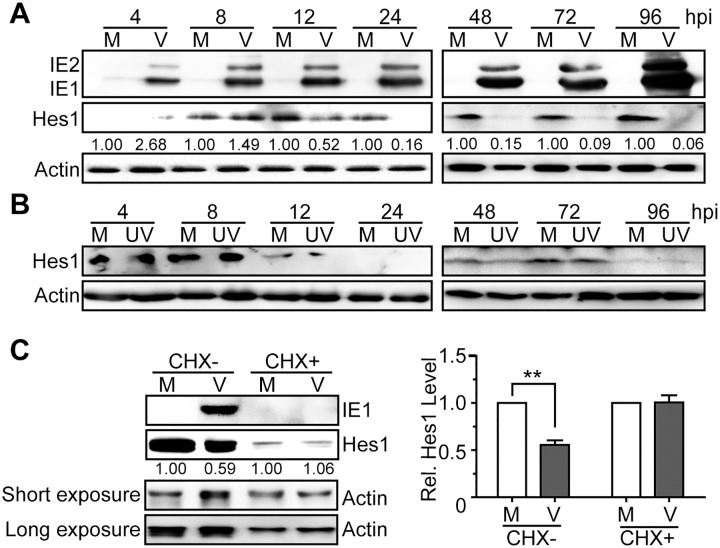
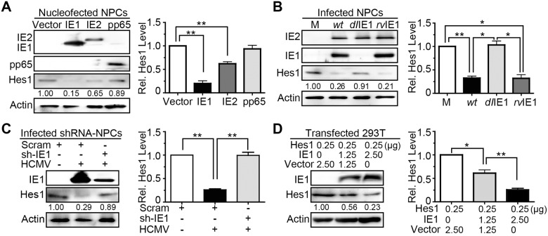
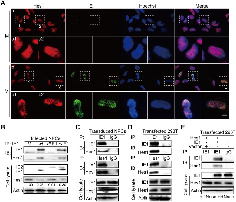
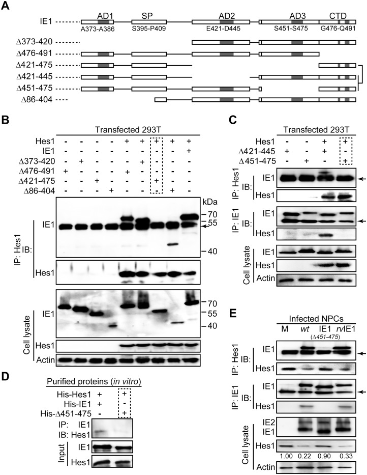
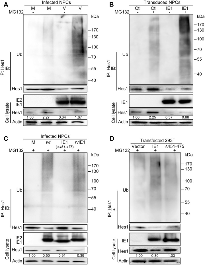
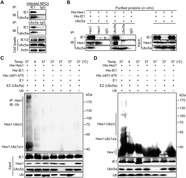
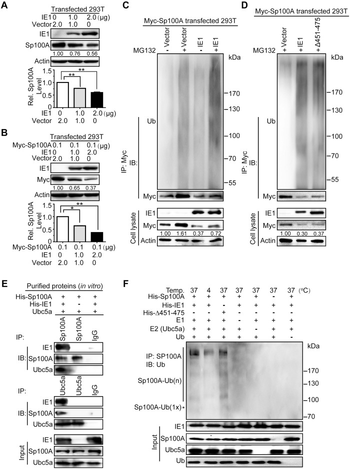
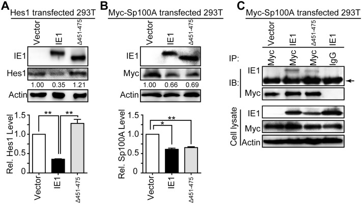
Congenital human cytomegalovirus (HCMV) infection is the leading cause of neurological disabilities in children worldwide, but the mechanisms underlying these disorders are far from well-defined. HCMV infection has been shown to dysregulate the Notch signaling pathway in human neural progenitor cells (NPCs). As an important downstream effector of Notch signaling, the transcriptional regulator Hairy and Enhancer of Split 1 (Hes1) is essential for governing NPC fate and fetal brain development. In the present study, we report that HCMV infection downregulates Hes1 protein levels in infected NPCs. The HCMV 72-kDa immediate-early 1 protein (IE1) is involved in Hes1 degradation by assembling a ubiquitination complex and promoting Hes1 ubiquitination as a potential E3 ubiquitin ligase, followed by proteasomal degradation of Hes1. Sp100A, an important component of PML nuclear bodies, is identified to be another target of IE1-mediated ubiquitination. A C-terminal acidic region in IE1, spanning amino acids 451 to 475, is required for IE1/Hes1 physical interaction and IE1-mediated Hes1 ubiquitination, but is dispensable for IE1/Sp100A interaction and ubiquitination. Our study suggests a novel mechanism linking downregulation of Hes1 protein to neurodevelopmental disorders caused by HCMV infection. Our findings also complement the current knowledge of herpesviruses by identifying IE1 as the first potential HCMV-encoded E3 ubiquitin ligase.


Conflict of interest statement

The authors have declared that no competing interests exist.

Figures

Fig 1
Fig 1. Downregulation of Hes1 protein during HCMV infection in NPCs requires de novo viral protein synthesis.
(A-B) Hes1 protein level in NPCs during HCMV infection. NPCs were mock-infected (M) or infected with HCMV (V) or UV-inactivated HCMV (UV) at an MOI of 3. Cells were harvested at the indicated times post infection, and subjected to immunoblotting (IB). The values listed below the blots indicate the relative Hes1 protein levels compared to corresponding mock controls following β-actin normalization. (C) Effect of HCMV infection on Hes1 protein levels following CHX treatment. NPCs nucleofected with 0.25μg pCDH-Hes1 were pre-treated with 0.1mg/ml CHX (CHX+) or equal volume of solvent control DMSO (CHX-) for 1h, followed by mock (M)- or HCMV(V)-infection (MOI = 5) in the presence of CHX or DMSO. Cells were collected at 12hpi and subjected to IB. The values listed below the blots indicate the relative Hes1 protein levels compared to corresponding mock controls following β-actin normalization. Representative images from 3 independent experiments are shown (left), and the relative levels of Hes1 are presented as the mean ± SD (right). **, P≤0.01.
Fig 2
Fig 2. HCMV IE1 is sufficient and necessary to reduce Hes1 protein levels.
(A) Effect of IE1, IE2 and pp65 expression on endogenous Hes1 protein levels in NPCs. At 48h following nucleofection with 5μg pCDH-GFP (vector), pCDH-IE1 (IE1), pCDH-IE2 (IE2) or pCDH-pp65 (pp65), NPCs were collected and subjected to IB for Hes1. (B) Effect of IE1 deletion on Hes1 protein downregulation during infection in NPCs. NPCs were mock-infected (M) or infected with TNwt (wt), TNdlIE1 (dlIE1) or TNrvIE1 (rvIE1) at an MOI of 10. Cells collected at 12hpi were subjected to IB. (C) Effect of IE1 knock-down on protein level of endogenous Hes1 during HCMV infection. At 48 h following transduction with lentiviruses expressing a scrambled shRNA (Scram) or an shRNA targeting IE1 (sh-IE1), NPCs were infected with HCMV (MOI = 1) for 24h and subjected to IB for Hes1 and IE1. (D) Effect of IE1 expression on protein levels of exogenous Hes1. At 48h following co-transfection with the indicated amount of pCDH-Hes1 (Hes1) and pEYFP-GFP (vector) or pEYFP-IE1 (IE1), 293T cells were collected and subjected to IB for Hes1. The values listed below the blots indicate the relative Hes1 protein levels compared to the corresponding controls following β-actin normalization. Representative images from 3 independent experiments are shown (left), and the relative levels of Hes1 are presented as the mean ± SD (right). *, P≤0.05; **, P≤0.01.
Fig 3
Fig 3. IE1 interacts with Hes1.
(A) Subcellular localization of IE1 and Hes1 in NPCs. Following mock (M) or HCMV infection (V) at an MOI of 0.5, NPCs grown on coverslips were collected at 4hpi for immunofluorescence assay. Shown are representative images from 3 independent experiments. The boxed regions are presented with higher magnification. Scale bar, 5μm. (B) Interaction of IE1 and Hes1 in HCMV infected NPCs. NPCs were harvested at 12hpi from mock (M), TNwt (wt), TNdlIE1 (dlIE1) or TNrvIE1 (rvIE1) infection (MOI = 10), and subjected to IE1-directed IP analysis and subsequent IB for Hes1 and IE1. The indicated proteins were also examined in cell lysates. The values listed below the blots indicate the relative protein level of Hes1 compared to the corresponding mock control following β-actin normalization. Representative results from 3 independent experiments are shown. (C) Interaction of IE1 and Hes1 in transduced NPCs. NPCs were transduced with lentivirus expressing IE1. Cells were harvested at 72 hour post transduction and subjected to either IE1- or Hes1-directed IP analysis and subsequent IB for Hes1 and IE1, with normal IgG as a nonspecific antibody control. Representative results from 3 independent experiments are shown. (D) IE1/Hes1 physical interaction in transfected 293T cells. 293T cells co-transfected with 6μg pCDH-Hes1 and 6μg pEYFP-IE1 were collected 48hpt. Cells were analyzed by IE1- or Hes1-directed IP (normal IgG served as a nonspecific antibody control) and subsequent IB for Hes1 and IE1, respectively. The total IE1 and Hes1 protein levels in cell lysates were also assessed. (E) IE1/Hes1 physical interaction in the absence of DNA and RNA. Following co-transfection with 6μg pCDH-Hes1 (Hes1) and 6μg pEYFP-GFP (vector) or pEYFP-IE1 (IE1) for 48h, 293T cells were harvested. Cell lysates were treated with DNase and RNase prior to IE1-directed IP (normal IgG served as a nonspecific antibody control) and subsequent IB for IE1 and Hes1. The total IE1 and Hes1 protein levels in cell lysates were also assessed.
Fig 4
Fig 4. The IE1/Hes1 interaction requires AA451-475 of IE1.
(A) Schematic diagram of wild-type IE1 and IE1 mutants used in this study. (B-C) IE1 region required for IE1/Hes1 interaction in vivo. Following co-transfection with 6μg plasmid pCDH-Hes1 and 6μg pEYFP -based plasmids expressing wild-type or mutant IE1 (Δ373–420, Δ476–491, Δ421–475, Δ86–404, Δ421–445 and Δ451–475) for 48h, 293T cells were sequentially processed for Hes1-directed IP and IB for IE1 and Hes1. The total IE1 and Hes1 protein levels in cell lysates were also assessed. Band corresponding to IgG heavy chains are indicated by arrows. (D) Interaction of purified IE1 or Δ451–475 and Hes1 in vitro. 1μg of each purified protein was mixed and subjected to pull down assay as described in Materials and Methods. (E) Interaction between IE1 or Δ451–475 and Hes1 in infected NPCs. Following mock (M), TNwt (wt), TN-IE1(Δ451–475) (IE1(Δ451–475)) or TNrvIE1 (rvIE1) infection at an MOI of 10, NPCs were harvested at 12hpi and cell lysates were subjected to either Hes1- or IE1-directed IP analysis and subsequent IB for IE1 or Hes1, respectively. The total IE1/2 and Hes1 protein levels in cell lysates were also assessed. The band corresponding to IgG heavy chain is indicated by an arrow.
Fig 5
Fig 5. IE1 prompts Hes1 ubiquitination in vivo.
(A) Effect of HCMV infection on ubiquitination of endogenous Hes1 in NPCs. Following mock (M) or HCMV infection (V) for 3h (MOI = 3), NPCs were treated with 12.5μM MG132 (+) or equivalent volume of DMSO (-) for 9h, and harvested at 12 hpi. Cell lysates were subjected to Hes1-directed IP and subsequent IB for ubiquitin (Ub) and Hes1. (B) Effect of IE1 on ubiquitination of endogenous Hes1 in transduced NPCs. NPCs were transduced with lentivirus expressing IE1 (IE1) or control (Ctl). Cells were treated with MG132 (+) or equivalent volume of DMSO (-) when GFP signal was clearly observed, harvested after being treated for 12 h, and subjected to Hes1-directed IP and subsequent IB for ubiquitin (Ub) and Hes1. (C) Effect of IE1 AA451-475 on ubiquitination of endogenous Hes1 in HCMV infected NPCs. NPCs were subjected to mock (M), TNwt (wt), TN-IE1(Δ451–475) (IE1(Δ451–475)) or TNrvIE1 (rvIE1) infection at an MOI of 10. At 3hpi, NPCs were treated with 12.5μM MG132 (+) for 9h, and then harvested at 12 hpi. Cell lysates were subjected to Hes1-directed IP and subsequent IB for ubiquitin (Ub) and Hes1. (D) Effect of IE1 AA451-475 on ubiquitination of exogenous Hes1 in 293T cells. 293T cells were co-transfected with 0.25μg pCDH-Hes1 and 2.5μg pEYFP (vector), pEYFP-IE1 (IE1) or pEYFP-IE1(Δ451–475) (Δ451–475). At 36 hpt, cells were treated with MG132 (+), and harvested after 12h treatment. Cell lysates were then subjected to Hes1-directed IP and subsequent IB for ubiquitin (Ub) and Hes1. For all the ubiquitination analysis in vivo, total levels of the indicated proteins were also examined in the corresponding cell lysates. The values listed below the Hes1 blots indicate the relative protein levels of Hes1 compared to the control(s) following β-actin normalization.
Fig 6
Fig 6. IE1 assembles a Hes1 ubiquitination complex and leads to ubiquitination of Hes1 in vitro.
(A) Interaction between IE1 and Ubc5a in HCMV infected NPCs. NPCs were infected with HCMV at an MOI of 3, and harvested at 12hpi. Cell lysates were subjected to IE1- or Ubc5a-directed IP followed by IB against IE1 and Ubc5a. Normal IgG was applied as the nonspecific antibody control. (B) Effect of IE1 on assembling the Hes1 and Ubc5a ubiquitination complex. The purified His-Hes1 (1μg) was mixed with Ubc5a (1μg) in the presence or absence of purified His-IE1 (1μg). The mixtures were then subjected to Hes1- or Ubc5a-directed IP, followed by IB for the indicated component proteins. Normal IgG was applied as the nonspecific antibody control. The input components in the reaction were examined. (C-D) Effect of IE1 AA451-475 on ubiquitination of Hes1 in vitro. Purified His-tagged proteins (His-Hes1, His-IE1, His-Δ451–475) were mixed with ubiquitin-activating enzyme (E1), ubiquitin-conjugating enzyme Ubc5a (E2) and ubiquitin as described in Materials and Methods, and then incubated at the indicated temperature for 2 h to perform the in vitro ubiquitination reaction. The mixtures were subjected to Hes1-directed IP and subsequent IB for ubiquitin (Ub) (C). Alternatively, the products from in vitro ubiquitination reactions were directly subjected to IB with anti-Hes1 (D). According to protein size, mono-ubiquitinated Hes1 is indicated by an asterisk (*). The input components in the reaction were examined, and the indicated proteins are shown.
Fig 7
Fig 7. IE1 induces ubiquitination of Sp100A and assembles an Sp100A ubiquitination complex.
(A-B) Effect of IE1 on the protein levels of endogenous and exogenous Sp100A in 293T cells. 293T cells were transfected with pEYFP-IE1 (IE1) or pEYFP (vector) in the absence (A) or presence (B) of pCMV-Myc-Sp100A (Myc-Sp100A). Cells were harvested at 48hpt for immunoblotting. Endogenous (A) or exogenous (B) Sp100A levels were examined by antibodies against Sp100A or Myc, respectively. The values listed below the blots indicate the relative protein levels of Sp100A compared to the controls following β-actin normalization. Representative images of IB from 3 independent experiments are shown (upper panel), and relative levels of Sp100A are presented as the mean ± SD (lower panel). **, P≤0.01. (C-D) Effect of IE1 on ubiquitination of exogenous Sp100A in 293T cells. 293T cells were transfected with 0.1μg pCMV-Myc-Sp100A (Myc-Sp100A) and 2μg pEYFP-IE1 (IE1), pEYFP-IE1(Δ451–475) (Δ451–475), or pEYFP (vector). At 36hpt, cells were treated with MG132 (+) or DMSO (-) for 12 h. Cell lysates were subjected to Myc-directed IP and subsequent IB for ubiquitin (Ub) and Myc. The indicated proteins were also examined in the cell lysates. (E) Effect of IE1 on assembling the Sp100A ubiquitination complex. Purified His-Sp100A was mixed with Ubc5a in the presence or absence of purified His-IE1. The mixtures were then subjected to Sp100A- or Ubc5a-directed IP, followed by IB for the indicated proteins. Representative results from 3 independent experiments are shown. Normal IgG was used as the nonspecific antibody control. The input components in the reaction were examined, and the indicated proteins are shown. (F) Effect of IE1 on ubiquitination of Sp100A in vitro. Purified His-IE1 or His-Δ451–475 were mixed together with His-Sp100A, E1, E2 (Ubc5a) and ubiquitin as described above and in Materials and Methods, and incubated at the indicated temperature for 2h to perform in vitro ubiquitination reaction. The mixtures were subjected to Sp100A-directed IP and subsequent IB for ubiquitin (Ub). The input components in the reaction were examined, and the indicated proteins are shown. The mono-ubiquitinated Sp100A is indicated by an asterisk (*) according to the protein size.
Fig 8
Fig 8. IE1 AA451-475 is dispensable for Sp100A interaction and downregulation.
(A-B) Effect of Δ451–475 on protein levels of exogenous Hes1 and Sp100A in 293T cells. 293T cells were co-transfected with 0.25μg pCDH-Hes1 (Hes1) (A) or pCMV-Myc-Sp100A (Myc-Sp100A) (B) along with 2.5μg pEYFP (vector), pEYFP-IE1 (IE1) or pEYFP-IE1(Δ451–475) (Δ451–475). Cells were harvested at 48hpt for IB to examine the protein levels of Hes1 (A) or Sp100A (B). The values listed below the blots indicate the relative protein levels of Hes1 or Sp100A compared to the controls following β-actin normalization. Representative images from 3 independent experiments are shown (upper panel), and relative protein levels are presented as the mean ± SD (lower panel). *, P≤0.05; **, P≤0.01. (C) Interaction of IE1 or Δ451–475 with Sp100A in 293T cells. 293T cells were transfected with 6μg pCMV-Myc-Sp100A (Myc-Sp100A) and 6μg pEYFP (vector), pEYFP-IE1 (IE1) or pEYFP-IE1(Δ451–475) (Δ451–475). Cells were harvested at 48hpt and cell lysates were subjected to Myc-directed IP and subsequent IB for IE1 and Myc. Normal IgG was applied as nonspecific antibody control, and the indicated proteins were examined in the cell lysates. The band corresponding to IgG heavy chain is indicated by an arrow.

Similar articles

Cited by

References

    1. Boppana SB, Pass RF, Britt WJ, Stagno S, Alford CA. Symptomatic Congenital Cytomegalovirus-Infection—Neonatal Morbidity and Mortality. Pediatr Infect Dis J. 1992;11(2):93–9. doi: 10.1097/00006454-199202000-00007 - DOI - PubMed
    1. Conboy TJ, Pass RF, Stagno S, Britt WJ, Alford CA, McFarland CE, et al. Intellectual development in school-aged children with asymptomatic congenital cytomegalovirus infection. Pediatrics. 1986;77(6):801–6. . - PubMed
    1. Rosenthal LS, Fowler KB, Boppana SB, Britt WJ, Pass RF, Schmid SD, et al. Cytomegalovirus Shedding and Delayed Sensorineural Hearing Loss Results From Longitudinal Follow-up of Children With Congenital Infection. Pediatr Infect Dis J. 2009;28(6):515–20. doi: 10.1097/INF.0b013e318198c724 - DOI - PMC - PubMed
    1. Stagno S, Pass RF, Cloud G, Britt WJ, Henderson RE, Walton PD, et al. Primary Cytomegalovirus-Infection in Pregnancy—Incidence, Transmission to Fetus, and Clinical Outcome. Jama-J Am Med Assoc. 1986;256(14):1904–8. - PubMed
    1. Li XJ, Liu XJ, Yang B, Fu YR, Zhao F, Shen ZZ, et al. Human Cytomegalovirus Infection Dysregulates the Localization and Stability of NICD1 and Jag1 in Neural Progenitor Cells. Journal of virology. 2015;89(13):6792–804. doi: 10.1128/JVI.00351-15 ; - DOI - PMC - PubMed

MeSH terms

Substances