. 2017 Jul 27;12(7):e0180869.
doi: 10.1371/journal.pone.0180869. eCollection 2017.

Roles of the low density lipoprotein receptor and related receptors in inhibition of lipoprotein(a) internalization by proprotein convertase subtilisin/kexin type 9

Affiliations

Roles of the low density lipoprotein receptor and related receptors in inhibition of lipoprotein(a) internalization by proprotein convertase subtilisin/kexin type 9

Rocco Romagnuolo et al. PLoS One. .

Abstract

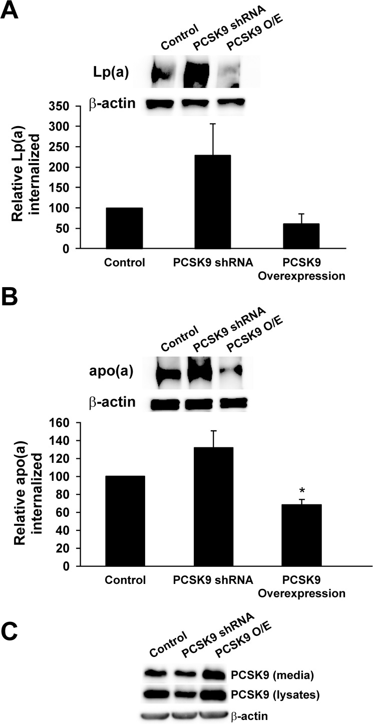
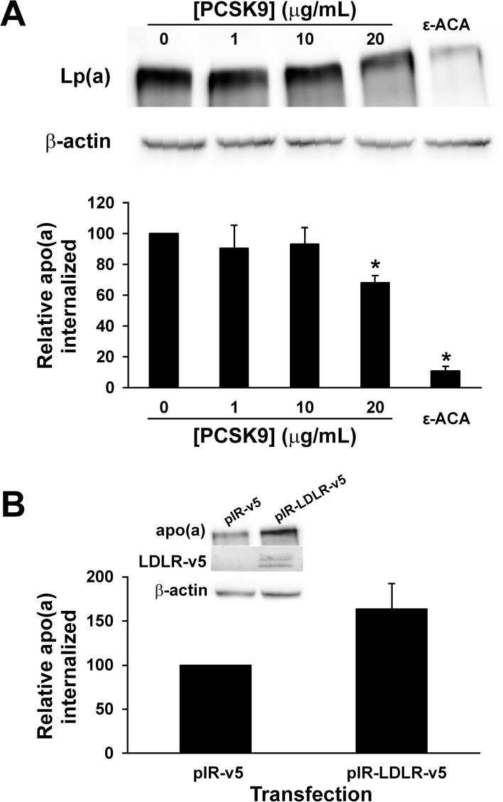
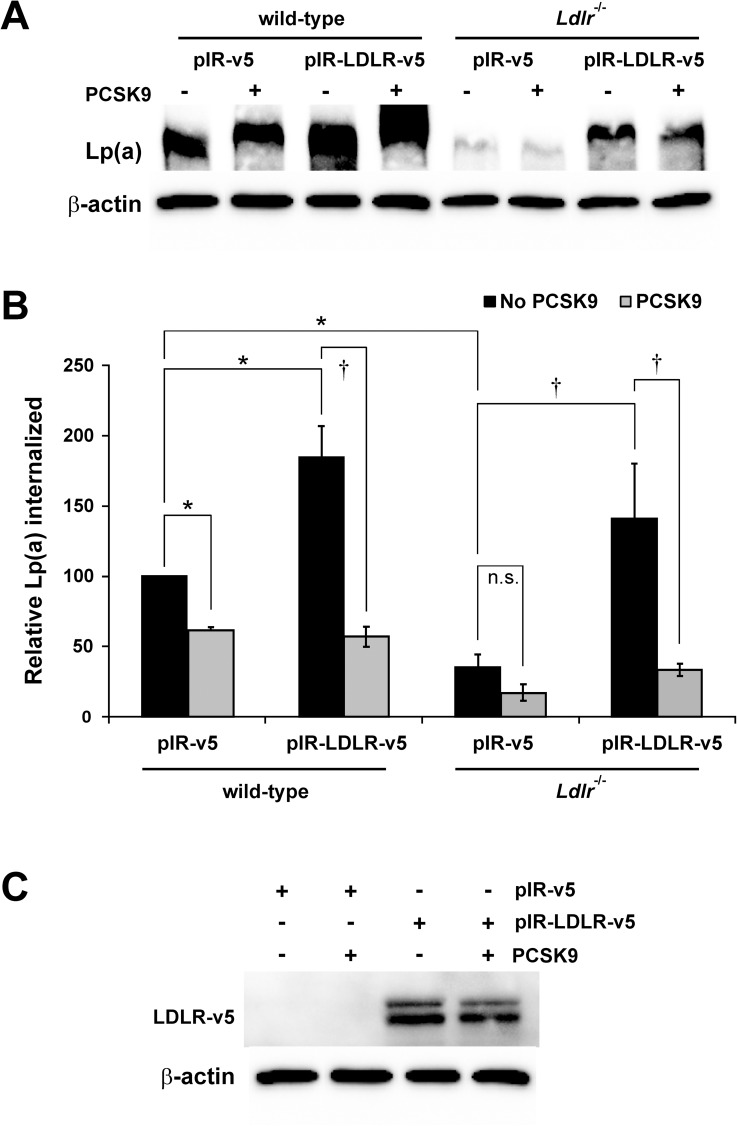
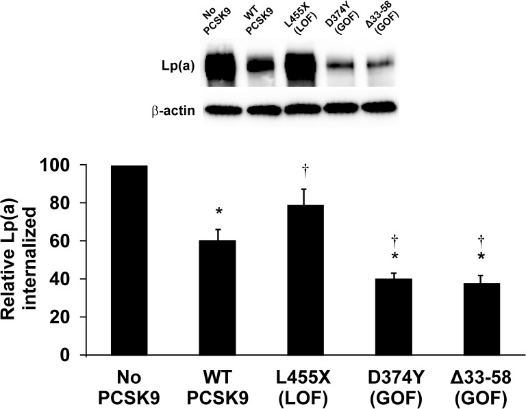
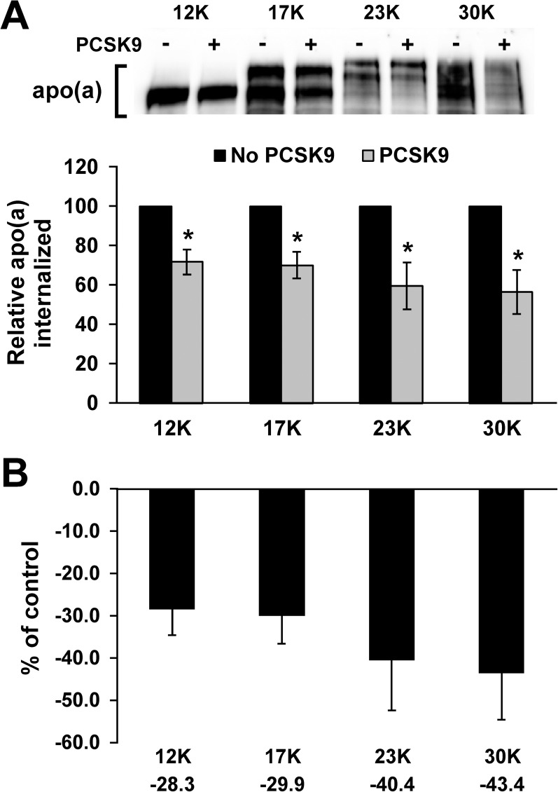
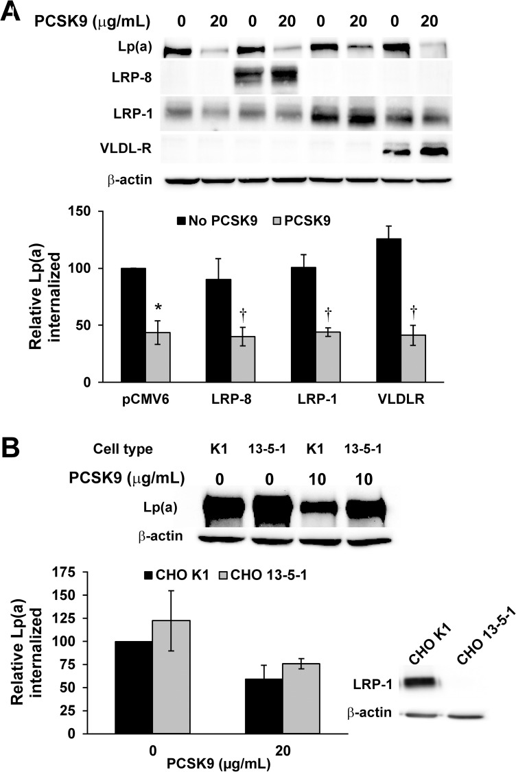
Elevated plasma concentrations of lipoprotein(a) (Lp(a)) are a causal risk factor for cardiovascular disease. The mechanisms underlying Lp(a) clearance from plasma remain unclear, which is an obvious barrier to the development of therapies to specifically lower levels of this lipoprotein. Recently, it has been documented that monoclonal antibody inhibitors of proprotein convertase subtilisin/kexin type 9 (PCSK9) can lower plasma Lp(a) levels by 30%. Since PCSK9 acts primarily through the low density lipoprotein receptor (LDLR), this result is in conflict with the prevailing view that the LDLR does not participate in Lp(a) clearance. To support our recent findings in HepG2 cells that the LDLR can act as a bona fide receptor for Lp(a) whose effects are sensitive to PCSK9, we undertook a series of Lp(a) internalization experiments using different hepatic cells, with different variants of PCSK9, and with different members of the LDLR family. We found that PCSK9 decreased Lp(a) and/or apo(a) internalization by Huh7 human hepatoma cells and by primary mouse and human hepatocytes. Overexpression of human LDLR appeared to enhance apo(a)/Lp(a) internalization in both types of primary cells. Importantly, internalization of Lp(a) by LDLR-deficient mouse hepatocytes was not affected by PCSK9, but the effect of PCSK9 was restored upon overexpression of human LDLR. In HepG2 cells, Lp(a) internalization was decreased by gain-of-function mutants of PCSK9 more than by wild-type PCSK9, and a loss-of function variant had a reduced ability to influence Lp(a) internalization. Apo(a) internalization by HepG2 cells was not affected by apo(a) isoform size. Finally, we showed that very low density lipoprotein receptor (VLDLR), LDR-related protein (LRP)-8, and LRP-1 do not play a role in Lp(a) internalization or the effect of PCSK9 on Lp(a) internalization. Our findings are consistent with the idea that PCSK9 inhibits Lp(a) clearance through the LDLR, but do not exclude other effects of PCSK9 such as on Lp(a) biosynthesis.


Conflict of interest statement

Competing Interests: M. Koschinsky has received research funding from Regeneron Pharmaceuticals, Inc. Ionis Pharmaceuticals, and Pfizer, Inc., is a member of an advisory board to Sanofi S.A., and has received speaker's honoraria from Regeneron Pharmaceuticals, Inc., Amgen, Inc., and Eli Lilly & Co., Ltd. M. Boffa has received research funding from Ionis Pharmaceuticals. Marcovina is a member of an advisory board to Sanofi S.A. The work was funded through a research contract from Regeneron Pharmaceuticals, Inc. The funders had no role in study design or data collection and analysis. The funders reviewed the manuscript and approved it for submission. This does not alter our adherence to PLOS ONE policies on sharing data and materials.

Figures

Fig 1
Fig 1. Internalization of Lp(a) by the Huh7 human hepatoma cell line.
Huh7 cells were stably transfected with expression plasmids encoding either PCSK9 or shRNA against PCSK9, or were mock-transfected. Cells were incubated with 10 μg/mL human Lp(a) (A) or 200 nM 17K apo(a) (B) for 4 hours. Cells were extensively washed to remove any bound Lp(a) and lysed to determine the relative amount of internalized Lp(a) compared to β-actin using western blot analysis. Representative blots are shown. The results represent the means ± s.e.m. of at least 3 independent experiments. *: p < 0.05 for indicated pairwise comparisons by one-sample t-test.
Fig 2
Fig 2. Internalization of apo(a) by primary human hepatocytes.
(A) Hepatocytes were plated on a collagen matrix and then incubated with 200 nM 17K apo(a) in the absence or presence of the indicated concentrations of PCSK9 or 200 mM ε-ACA for 4 hours. Cells were extensively washed to remove any bound apo(a) and lysed to determine the relative amount of internalized apo(a) compared to β-actin using western blot analysis. Representative blots are also shown. The results represent the means ± s.e.m. of at least 3 independent experiments. *: p < 0.05 for indicated pairwise comparisons by one-sample t-test. (B) Hepatocytes were transiently transfected with an expression vector encoding human LDLR (pIR-LDLR-v5) or the corresponding empty expression vector. Internalization of 17K apo(a) was determined as described for Panel A. Also shown are representative blots probed with an anti-apo(a) antibody for apo(a) internalization and an anti-v5 antibody for LDLR overexpression.
Fig 3
Fig 3. Internalization of Lp(a) by primary mouse hepatocytes.
Hepatocytes were isolated from wild-type (left) and Ldlr-/- (right) mice. Where indicated, cells were transiently transfected with an expression vector encoding human LDLR (pIR-LDLR-v5) or the corresponding empty expression vector (pIR-v5). Cells were incubated with 10 μg/mL Lp(a) in the absence or presence of 20 μg/mL PCSK9 for 4 hours. Cells were extensively washed to remove any bound Lp(a) and lysed to determine the relative amount of internalized Lp(a) compared to β-actin using western blot analysis. (A) Representative blots are shown. (B) graphical representation of the results, representing the means ± s.e.m. of at least 3 independent experiments. *: p < 0.05 versus control by one-sample t-test; †: p < 0.05 for indicated pairwise comparison by Student’s t-test; n.s.: not significant. (C) Representative western blot (from wild-type hepatocytes) showing overexpression of LDLR in the presence or absence of added PCSK9 using an anti-v5 antibody.
Fig 4
Fig 4. Effect of GOF and LOF variants of PCSK9 on Lp(a) internalization.
HepG2 cells were treated with either no PCSK9, or 10 μg/mL purified PCSK9 variants. Cells were incubated with 10 μg/mL purified human Lp(a) for 4 hours. Cells were extensively washed to remove any bound Lp(a) and lysed to determine the relative amount of internalized Lp(a) compared to β-actin using western blot analysis. Representative blots are shown. The data represent the means ± s.e.m. of at least 4 independent experiments. *: p < 0.05 vs absence of PCSK9 by one-sample t-test; †: p < 0.05 versus wild-type (WT) PCSK9 by Student’s t-test.
Fig 5
Fig 5. Effect of apo(a) isoform size on the ability of PCSK9 to regulate internalization.
(A) HepG2 cells were treated with the indicated recombinant apo(a) variants (200 nM) in the presence or absence of 10 μg/mL purified PCSK9 for 4 hours. Cells were extensively washed to remove any bound apo(a) and lysed to determine the relative amount of internalized apo(a) compared to β-actin using western blot analysis. The internalization values in the presence of PCSK9 are expressed relative to the values obtained for that particular isoform in the absence of PCSK9. Representative blots are shown. The data represent the means ± s.e.m. of at least 7 independent experiments. *: p < 0.05 vs absence of PCSK9 by Student’s t-test. (B) The percent decrease in apo(a) internalization evoked by PCSK9 was calculated from the data in (A) and is plotted for each apo(a) isoform. No significant differences were observed (by one-way ANOVA).
Fig 6
Fig 6. Role of LDLR-related receptors in Lp(a) internalization.
(A) HepG2 cells were transiently transfected with the indicated expression vectors or the empty parental pCMV6 vector. Cells were incubated with 10 μg/mL purified human Lp(a) for 4 hours. Cells were extensively washed to remove any bound Lp(a) and lysed to determine the relative amount of internalized Lp(a) compared to β-actin using western blot analysis. Representative western blots for Lp(a), β-actin, and the respective ectopically-expressed receptors are shown. (B) Lp(a) internalization assays were performed as in Panel A, except in the LRP-1-expressing CHO cell line K1 or the LRP-deficient CHO cell line 13-5-1. Also shown is a western blot confirming the absence of LRP-1 in the 13-5-1 cell line. The data represent the means ± s.e.m. of at least 3 independent experiments. *: p < 0.05 vs absence of PCSK9 by one-sample t-test; †: p < 0.05 versus absence of PCSK9 by Student’s t-test.

Similar articles

Cited by

References

    1. Clarke R, Peden JF, Hopewell JC, Kyriakou T, Goel A, Heath SC, et al. Genetic variants associated with Lp(a) lipoprotein level and coronary disease. N Engl J Med. 2009;361(26):2518–28. doi: 10.1056/NEJMoa0902604 . - DOI - PubMed
    1. Kamstrup PR, Tybjaerg-Hansen A, Steffensen R, Nordestgaard BG. Genetically elevated lipoprotein(a) and increased risk of myocardial infarction. JAMA. 2009;301(22):2331–9. doi: 10.1001/jama.2009.801 . - DOI - PubMed
    1. Koschinsky ML, Boffa MB. Lipoprotein(a): an important cardiovascular risk factor and a clinical conundrum. Endocrinol Metab Clin North Am. 2014;43(4):949–62. doi: 10.1016/j.ecl.2014.08.002 . - DOI - PubMed
    1. McLean JW, Tomlinson JE, Kuang WJ, Eaton DL, Chen EY, Fless GM, et al. cDNA sequence of human apolipoprotein(a) is homologous to plasminogen. Nature. 1987;330(6144):132–7. doi: 10.1038/330132a0 . - DOI - PubMed
    1. van der Hoek YY, Wittekoek ME, Beisiegel U, Kastelein JJ, Koschinsky ML. The apolipoprotein(a) kringle IV repeats which differ from the major repeat kringle are present in variably-sized isoforms. Hum Mol Genet. 1993;2(4):361–6. . - PubMed

LinkOut - more resources