

Magnetite-Quantum Dot Immunoarray for Plasmon-Coupled-Fluorescence Imaging of Blood Insulin and Glycated Hemoglobin
- PMID: 28750536
- PMCID: PMC5931802
- DOI: 10.1021/acssensors.7b00124
Magnetite-Quantum Dot Immunoarray for Plasmon-Coupled-Fluorescence Imaging of Blood Insulin and Glycated Hemoglobin
Abstract
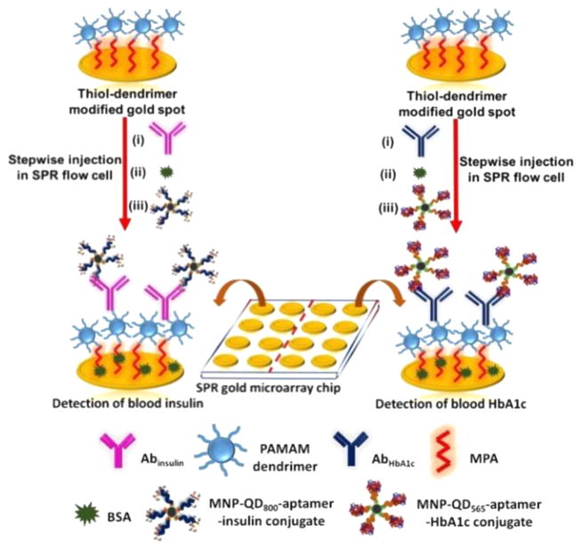
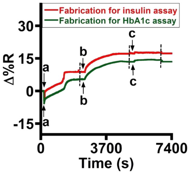
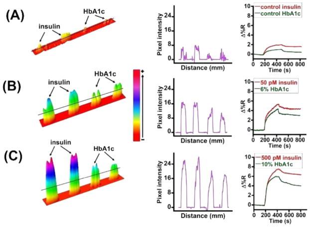
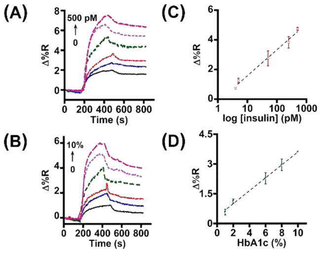
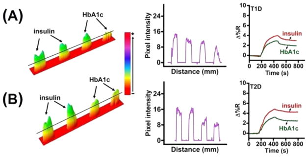
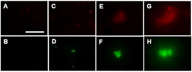
New microarray chip strategies that are sensitive and selective and that can measure low levels of important biomarkers directly in a blood sample are significant for improving human health by allowing timely diagnosis of an abnormal condition. Herein, we designed an antibody-aptamer immunoarray chip to demonstrate simultaneous measurement of blood insulin and glycated hemoglobin (HbA1c) levels relevant to diabetic and prediabetic disorders using a surface plasmon microarray with validation by fluorescence imaging. To accomplish both surface plasmon and fluorescence imaging on the same sample, we decorated magnetite nanoparticles with quantum dots for covalent immobilization of aptamers for subsequent capture and isolation of the aptamers specific for insulin and HbA1c markers from 20-times diluted whole blood samples. Direct clinically relevant analysis, along with fluorescent imaging of the two markers, was achieved by this new immunoarray platform. The limit of detection was 4 pM for insulin and 1% for HbA1c. Examination of cross-talk using thrombin and platelet-derived growth factor confirmed that the designed immunoarray was highly selective for insulin and HbA1c. Surface plasmon kinetic analysis provided apparent binding constants of 0.24 (±0.08) nM and 37 (±3) μM, respectively, for the binding of insulin and HbA1c onto their surface immobilized monoclonal antibodies. Thus, quantitative imaging of ultralow levels of blood biomarker levels with binding kinetics is uniquely obtained in the designed immunoarray chip. In conclusion, this report demonstrates considerable significance of the developed magnetite-quantum dot-bioconjugate strategy for clinical diagnostics of whole blood biomarkers with characterization of molecular binding interactions.
Keywords: binding kinetics; magnetite-quantum dot; microarray; nonglucose markers; plasmon-coupled-fluorescence; whole blood analysis.
Conflict of interest statement
The authors declare no competing financial interest.
Figures
Similar articles
-
Magnetic Optical Microarray Imager for Diagnosing Type of Diabetes in Clinical Blood Serum Samples.ACS Sens. 2016 Apr 22;1(4):437-443. doi: 10.1021/acssensors.5b00273. Epub 2016 Feb 18. ACS Sens. 2016. PMID: 27231720 Free PMC article.
-
Ultrasensitive quantum dot-coupled-surface plasmon microfluidic aptasensor array for serum insulin detection.Talanta. 2020 Nov 1;219:121314. doi: 10.1016/j.talanta.2020.121314. Epub 2020 Jun 30. Talanta. 2020. PMID: 32887054
-
Single-step bioassays in serum and whole blood with a smartphone, quantum dots and paper-in-PDMS chips.Analyst. 2015 Jun 21;140(12):4037-45. doi: 10.1039/c5an00475f. Epub 2015 Apr 30. Analyst. 2015. PMID: 25924885
-
Compact quantitative optic fiber-based immunoarray biosensor for rapid detection of small analytes.Biosens Bioelectron. 2010 Sep 15;26(1):16-22. doi: 10.1016/j.bios.2010.04.044. Epub 2010 May 7. Biosens Bioelectron. 2010. PMID: 20837444
-
Aptamers-based sandwich assay for silver-enhanced fluorescence multiplex detection.Anal Chim Acta. 2016 Jan 28;905:149-55. doi: 10.1016/j.aca.2015.12.013. Epub 2015 Dec 17. Anal Chim Acta. 2016. PMID: 26755149
Cited by
-
Epitope-Resolved Detection of Peanut-Specific IgE Antibodies by Surface Plasmon Resonance Imaging.Chembiochem. 2018 Feb 2;19(3):199-202. doi: 10.1002/cbic.201700513. Epub 2018 Jan 4. Chembiochem. 2018. PMID: 29232483 Free PMC article.
-
Advances in Nanomaterial-based Biosensors for Determination of Glycated Hemoglobin.Curr Top Med Chem. 2022;22(27):2261-2281. doi: 10.2174/1568026622666220915114646. Curr Top Med Chem. 2022. PMID: 36111762
-
Emerging biosensor probes for glycated hemoglobin (HbA1c) detection.Mikrochim Acta. 2024 May 6;191(6):300. doi: 10.1007/s00604-024-06380-7. Mikrochim Acta. 2024. PMID: 38709399 Review.
-
Building Biomimetic Potency Tests for Islet Transplantation.Diabetes. 2021 Feb;70(2):347-363. doi: 10.2337/db20-0297. Diabetes. 2021. PMID: 33472944 Free PMC article. Review.
-
Emerging Theranostic Nanomaterials in Diabetes and Its Complications.Adv Sci (Weinh). 2022 Jan;9(3):e2102466. doi: 10.1002/advs.202102466. Epub 2021 Nov 25. Adv Sci (Weinh). 2022. PMID: 34825525 Free PMC article. Review.
References
-
- Centers for Disease Control and Prevention. National Diabetes Fact Sheet. 2011 http://www.cdc.gov/diabetes/pubs/pdf/ndfs_2011.pdf.
-
- Pribyl J, Skladal P. Development of a combined setup for simultaneous detection of total and glycated haemoglobin content in blood samples. Biosens Bioelectron. 2006;21:1952–1959. - PubMed
-
- Gallagher EJ, Le Roith D, Bloomgarden Z. Review of hemoglobin A(1c) in the management of diabetes. J Diabetes. 2009;1:9–17. - PubMed
-
- Wareham NJ, Pfister R. Diabetes: glycated hemoglobin is a marker of diabetes and CVD risk. Nat Rev Cardiol. 2010;7:367–368. - PubMed
Grants and funding
LinkOut - more resources
Full Text Sources
Other Literature Sources
