. 2017 Jul 27;19(1):54.
doi: 10.1186/s12968-017-0366-2.

CMR fluoroscopy right heart catheterization for cardiac output and pulmonary vascular resistance: results in 102 patients

Affiliations

CMR fluoroscopy right heart catheterization for cardiac output and pulmonary vascular resistance: results in 102 patients

Toby Rogers et al. J Cardiovasc Magn Reson. .

Abstract

Background: Quantification of cardiac output and pulmonary vascular resistance (PVR) are critical components of invasive hemodynamic assessment, and can be measured concurrently with pressures using phase contrast CMR flow during real-time CMR guided cardiac catheterization.

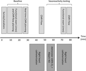
Methods: One hundred two consecutive patients underwent CMR fluoroscopy guided right heart catheterization (RHC) with simultaneous measurement of pressure, cardiac output and pulmonary vascular resistance using CMR flow and the Fick principle for comparison. Procedural success, catheterization time and adverse events were prospectively collected.

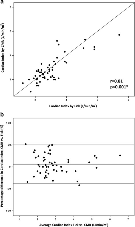
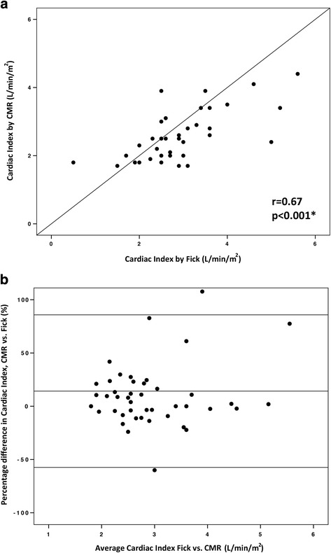
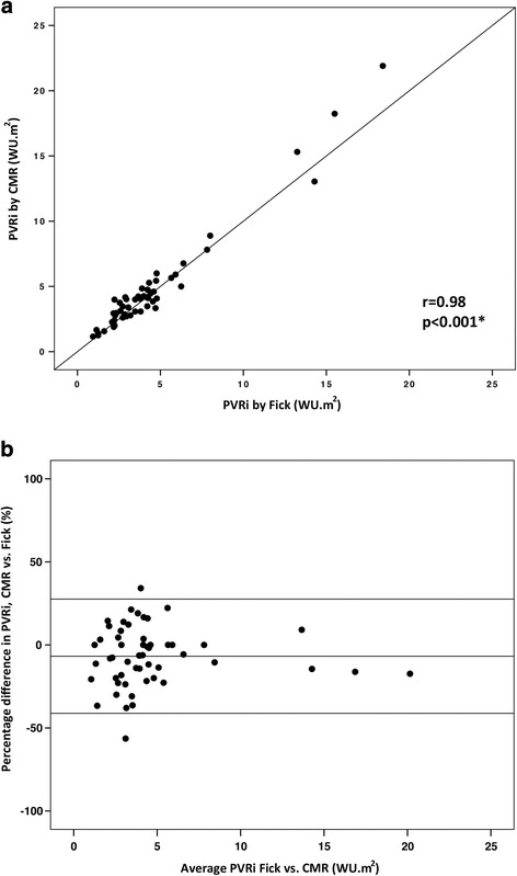
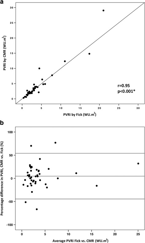
Results: RHC was successfully completed in 97/102 (95.1%) patients without complication. Catheterization time was 20 ± 11 min. In patients with and without pulmonary hypertension, baseline mean pulmonary artery pressure was 39 ± 12 mmHg vs. 18 ± 4 mmHg (p < 0.001), right ventricular (RV) end diastolic volume was 104 ± 64 vs. 74 ± 24 (p = 0.02), and RV end-systolic volume was 49 ± 30 vs. 31 ± 13 (p = 0.004) respectively. 103 paired cardiac output and 99 paired PVR calculations across multiple conditions were analyzed. At baseline, the bias between cardiac output by CMR and Fick was 5.9% with limits of agreement -38.3% and 50.2% with r = 0.81 (p < 0.001). The bias between PVR by CMR and Fick was -0.02 WU.m2 with limits of agreement -2.6 and 2.5 WU.m2 with r = 0.98 (p < 0.001). Correlation coefficients were lower and limits of agreement wider during physiological provocation with inhaled 100% oxygen and 40 ppm nitric oxide.

Conclusions: CMR fluoroscopy guided cardiac catheterization is safe, with acceptable procedure times and high procedural success rate. Cardiac output and PVR measurements using CMR flow correlated well with the Fick at baseline and are likely more accurate during physiological provocation with supplemental high-concentration inhaled oxygen.

Trial registration: Clinicaltrials.gov NCT01287026 , registered January 25, 2011.

Keywords: CMR; Cardiac MRI; Interventional MRI catheterization; Invasive hemodynamics; Phase contrast MRI flow; Real-time MRI; Right heart catheterization.


Conflict of interest statement

Authors’ information

Not applicable.

Ethics approval and consent to participate

The study was approved by the NHLBI Institutional Review Board and conducted under NHLBI Federalwide Assurance number FWA00000004. All subjects provided written informed consent.

Consent for publication

The protocol and NHLBI IRB approval include permission to use de-identified subject images in scientific communications.

Competing interests

NIH and Siemens Medical Systems have a collaborative research and development agreement for interventional CMR.

Publisher’s Note

Springer Nature remains neutral with regard to jurisdictional claims in published maps and institutional affiliations.

Figures

Fig. 1
Fig. 1
Interventional CMR (iCMR) cardiac catheterization laboratory. a Vascular access is obtained in the X-ray room. b The patient is transferred from X-ray to CMR using a transfer table that docks with the CMR table. Hemodynamic monitoring is seamless between the two modalities. c View of the CMR room from the X-ray side of the iCMR laboratory. d Transfemoral cardiac catheterization under CMR guidance. Real-time images are projected in the CMR room for the operator to view. Noise-cancelling headsets permit open communication with the iCMR control room
Fig. 2
Fig. 2
Example CMR-safe catheters for right heart catheterization. A selection of different shape and stiffness CMR-safe balloon catheters is useful to navigate different right heart anatomies. a Vascor Balloon Wedge Pressure Catheter, Model #172-110P; (b) Medtronic Pulmonary Wedge Pressure Catheter, Model #150075; (c) Edwards True Size Monitoring ‘S’ Tip Catheter, Model #S111F7
Fig. 3
Fig. 3
CMR fluoroscopy guided right heart catheterization. a Coronal view with the gadolinium filled balloon at the tip of the catheter in the inferior vena cava (arrow), (b) sagittal view of the superior vena cava, (c) coronal view of the right ventricle, and (d) axial view of the main pulmonary artery bifurcation with the balloon in the right pulmonary artery
Fig. 4
Fig. 4
Procedure workflow for CMR guided right heart catheterization
Fig. 5
Fig. 5
Comparison of cardiac index calculated with the Fick principle and with CMR flow at baseline on room air. a Correlation between cardiac index calculated with the Fick principle and with CMR flow at baseline on room air (n = 56 paired calculations). b Bland-Altman plot of the difference in cardiac index calculated with the two methods, and mean cardiac index with the two methods
Fig. 6
Fig. 6
Comparison of cardiac index calculated with the Fick principle and with CMR flow on inhaled 100% O2 and 40 ppm NO. a Correlation between cardiac index calculated with the Fick principle and with CMR flow on inhaled 100% O2 and 40 ppm NO (n = 47 paired calculations). b Bland-Altman plot of the difference in cardiac index calculated with the two methods, and mean cardiac index with the two methods
Fig. 7
Fig. 7
Comparison of PVRi calculated with the Fick principle and with CMR flow at baseline on room air. a Correlation between PVRi calculated with the Fick principle and with CMR flow at baseline on room air (n = 54 paired calculations). b Bland-Altman plot of the difference in PVRi calculated with the two methods, and mean PVRi with the two methods. PVRi: pulmonary vascular resistance index
Fig. 8
Fig. 8
Comparison of PVRi calculated with the Fick principle and with CMR flow on inhaled 100% O2 and 40 ppm NO. a Correlation between PVRi calculated with the Fick principle and with CMR flow on inhaled 100% O2 and 40 ppm NO (n = 45 paired calculations). b Bland-Altman plot of the difference in PVRi calculated with the two methods, and mean PVRi with the two methods. PVRi: pulmonary vascular resistance index

References

    1. Winniford MD, Kern MJ, Lambert CR. Blood flow measurement and quantification of vascular stenoses. In: Pepine CJ, Hill JA, Lambert CR, editors. Diagnostic and therapeutic cardiac catheterization 3/e. Baltimore: Williams & Wilkins; 1998. pp. 399–441.
    1. LaFarge CG, Miettinen OS. The estimation of oxygen consumption. Cardiovasc Res. 1970;4:23–30. doi: 10.1093/cvr/4.1.23. - DOI - PubMed
    1. Dehmer GJ, Firth BG, Hillis LD. Oxygen consumption in adult patients during cardiac catheterization. Clin Cardiol. 1982;5:436–440. doi: 10.1002/clc.4960050803. - DOI - PubMed
    1. Narang N, Thibodeau JT, Levine BD, Gore MO, Ayers CR, Lange RA, et al. Inaccuracy of estimated resting oxygen uptake in the clinical setting. Circulation. 2014;129:203–210. doi: 10.1161/CIRCULATIONAHA.113.003334. - DOI - PubMed
    1. Nayak KS, Nielsen JF, Bernstein MA, Markl M, Gatehouse PD, Botnar RM, et al. Cardiovascular magnetic resonance phase contrast imaging. J Cardiovasc Magn Reson. 2015;17:71. doi: 10.1186/s12968-015-0172-7. - DOI - PMC - PubMed

MeSH terms

Associated data