. 2017 Jul 27;18(1):140.
doi: 10.1186/s13059-017-1267-2.

ReMixT: clone-specific genomic structure estimation in cancer

Affiliations

ReMixT: clone-specific genomic structure estimation in cancer

Andrew W McPherson et al. Genome Biol. .

Erratum in

Abstract

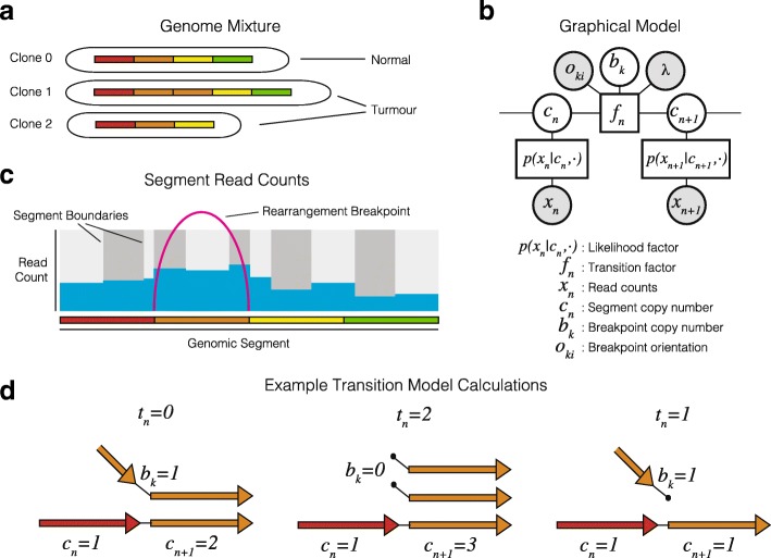
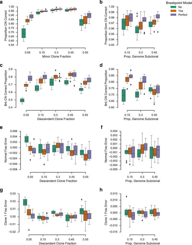
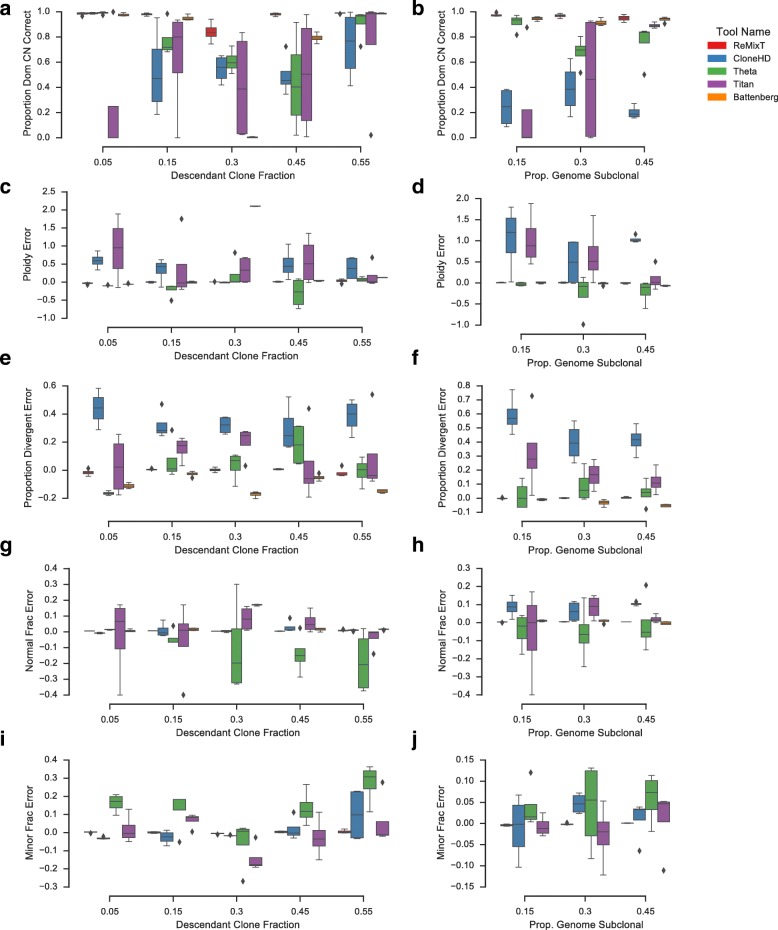
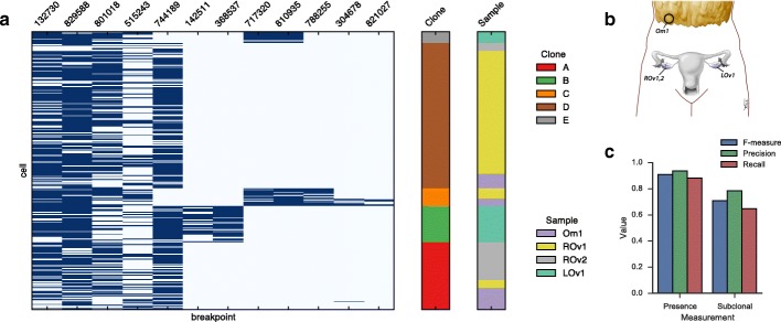
Somatic evolution of malignant cells produces tumors composed of multiple clonal populations, distinguished in part by rearrangements and copy number changes affecting chromosomal segments. Whole genome sequencing mixes the signals of sampled populations, diluting the signals of clone-specific aberrations, and complicating estimation of clone-specific genotypes. We introduce ReMixT, a method to unmix tumor and contaminating normal signals and jointly predict mixture proportions, clone-specific segment copy number, and clone specificity of breakpoints. ReMixT is free, open-source software and is available at http://bitbucket.org/dranew/remixt .

Keywords: Cancer genomics; Copy number variation; DNA sequencing; Genomic rearrangement; Tumour heterogeneity.


Conflict of interest statement

Ethics approval and consent to participate

High-grade serous ovarian cancer.

Ethical approval was obtained from the University of British Columbia (UBC) Research Ethics Board (H08-01411 NGS Huntsman). Women undergoing debulking surgery (primary or recurrent) for carcinoma of ovarian, peritoneal, and/or fallopian tube origin were approached for informed consent for the banking of tumour tissue. All experimental methods comply with the Helsinki Declaration.

Breast cancer

Anonymized tumour tissue from women aged 26–82 undergoing surgery or diagnostic core biopsy was collected with informed consent, according to procedures approved by the UBC Research Ethics Board (H06-00289 Breast Tumour Tissue Repository and H13-01125 Breast Xenograft Aparicio). All experimental methods comply with the Helsinki Declaration.

Patient-derived xenografts

Female NOD/SCID interleukin-2 receptor gamma null (NSG) and NOD Rag-1 null interleukin-2 receptor gamma null (NRG) mice were bred and housed at the Animal Resource Centre at the British Columbia Cancer Research Centre and the Biological Resource Unit at the Cancer Research UK Cambridge Research Institute. Surgery was carried out on mice between the ages of 5–10 weeks. All experimental procedures were approved by the University of British Columbia Animal Care Committee and the University of Cambridge Animal Welfare and Ethical Review Committee.

Consent for publication

Not applicable.

Competing interests

The authors declare that they have no competing interests.

Publisher’s Note

Springer Nature remains neutral with regard to jurisdictional claims in published maps and institutional affiliations.

Figures

Fig. 1
Fig. 1
An overview of the ReMixT Method. a) Bulk sequencing is applied to a mixture of cells modeled as a set of clones of unknown proportion each with distinct sets of chromosomes with unknown structure. b) Observed data include binned read counts per segment, and rearrangement breakpoints connecting segment ends. c) The ReMixT graphical model as a factor graph. d) Calculation of the transition factor involves calculating the number of telomeres t, the number of segment ends left unconnected to another segment end in the model
Fig. 2
Fig. 2
Simulation results for the integrated breakpoint model and an equivalent hidden Markov model (HMM) with postprocessing to infer breakpoint copy number. Also shown are results for the breakpoint model with perfect initialization. Two sets of simulations were performed, varying fraction of the descendant tumour clone (left column) and proportion of the genome with divergent copy number (right column). Boxplots show proportion of the genome (a, b) and proportion of breakpoints (c, d) for which the tool correctly called clone-specific copy number, in addition to relative normal fraction error (e, f) and relative minor clone fraction error (g, h). Boxes show the interquartile (IQR) range with a line depicting the median. Whiskers extend 1.5×IQR above quartile 3 and below quartile 1. Diamonds show positions of outlier data points
Fig. 3
Fig. 3
Performance comparison of ReMixT with CloneHD, TITAN, Battenberg, and THetA using read re-sampling simulations. Two sets of simulations were performed, varying fraction of the descendant tumour clone (left column) and proportion of the genome with divergent copy number (right column). Boxplots show proportion of the genome for which the tool correctly called the copy number of the dominant clone (a, b), relative mean ploidy error compared to simulated (c, d), relative proportion divergent error compared to simulated (e, f), relative normal fraction estimation error compared to simulated (g, h), and relative minor clone fraction estimation error compared to simulated (i, j). Battenberg was excluded from the minor clone fraction benchmark, as it does not produce a global estimate of this parameter. Boxes show the interquartile (IQR) range with a line depicting the median. Whiskers extend 1.5×IQR above quartile 3 and below quartile 1. Diamonds show positions of outlier data points
Fig. 4
Fig. 4
Single cell validation of ReMixT results for 12 breakpoints in 294 cells from 4 HGS Ovarian tumour samples: Omentum 1 (Om1), Right Ovary 1 and 2 (ROv1 and ROv2), and Left Ovary 1 (LOv1). (a) Breakpoint (x-axis) by cell (y-axis) presence (dark blue) / absence (light blue) with cells annotated by sample of origin and clone as inferred by the Single Cell Genotyper. (b) Approximate anatomic location of the 4 tumour samples. (c) F-measure, precision and recall for ReMixT calls of breakpoint presence and subclonality
Fig. 5
Fig. 5
Tracking clonal expansions in xenograft passages. a Breakpoints identified by ReMixT as clone-specific were classified according to their clonal prevalence change between SA501X1A and replicate xenograft passages SA501X3A and SA501X3F. All breakpoints could be classified as ascending in both SA501X3A and SA501X3F, descending in both, or stable in at least one. Shown are the clonal prevalence changes between pairs of samples for which WGS was available. b Relationship between primary tumour sample T and xenograft passages X*. c Accuracy of copy number inference for X3F based on single cell whole genome sequencing. Shown is the proportion of regions with correctly predicted copy number (y-axis) for each clone A copy number (x-axis), split between clonal and subclonal (blue/green) as determined from single cell data. d Copy number profile (top) for chromosomes 7 and 15 showing corroboration between single cell (bottom) and ReMixT (middle) subclonal copy number prediction. Yellow flags show the location of translocation breakpoints predicted to be subclonal by ReMixT. e Similarly, chromosomes 1/18 translocation breakpoints predicted to be subclonal by ReMixT. Copy number plots show raw major (red) and minor (blue) copy numbers
Fig. 6
Fig. 6
Inference of partial tumour chromosome assemblies based on linking subclonal segments and breakpoints. Two assembled chromosomes are shown for cell lines DAH354 (a) and DAH355 (b). Shown for each assembled chromosome is a schematic of the segments involved (top left), a table of breakpoint copy number predicted by ReMixT (top right), and a chromosome copy number plot (bottom). Each copy number plot shows raw major (red) and minor (blue) copy numbers (top axis), in addition to prediction of subclonality (bottom axis)

References

    1. Rausch T, Zichner T, Schlattl A, Stütz AM, Benes V, Korbel JO. Delly: structural variant discovery by integrated paired-end and split-read analysis. Bioinformatics. 2012;28(18):333–9. doi: 10.1093/bioinformatics/bts378. - DOI - PMC - PubMed
    1. Hormozdiari F, Alkan C, Eichler EE, Sahinalp SC. Combinatorial algorithms for structural variation detection in high-throughput sequenced genomes. Genome Res. 2009;19(7):1270–8. doi: 10.1101/gr.088633.108. - DOI - PMC - PubMed
    1. Layer RM, Chiang C, Quinlan AR, Hall IM. Lumpy: a probabilistic framework for structural variant discovery. Genome Biol. 2014;15(6):84. doi: 10.1186/gb-2014-15-6-r84. - DOI - PMC - PubMed
    1. McPherson A, Shah SP, Sahinalp SC. deStruct: accurate rearrangement detection using breakpoint specific realignment. bioRxiv. 2017. https://doi.org/10.1101/117523. - DOI
    1. Ha G, Roth A, Khattra J, Ho J, Yap D, Prentice LM, Melnyk N, McPherson A, Bashashati A, Laks E, Biele J, Ding J, Le A, Rosner J, Shumansky K, Marra MA, Gilks CB, Huntsman DG, McAlpine JN, Aparicio S, Shah SP. TITAN: inference of copy number architectures in clonal cell populations from tumor whole-genome sequence data. Genome Res. 2014;24(11):1881–93. doi: 10.1101/gr.180281.114. - DOI - PMC - PubMed

Publication types

MeSH terms