Comparative Study
. 2017 Sep 22;292(38):15870-15879.
doi: 10.1074/jbc.M117.804625. Epub 2017 Jul 27.

Kinetic analyses of single-stranded break repair by human DNA ligase III isoforms reveal biochemical differences from DNA ligase I

Affiliations
Comparative Study

Kinetic analyses of single-stranded break repair by human DNA ligase III isoforms reveal biochemical differences from DNA ligase I

Justin R McNally et al. J Biol Chem. .

Abstract

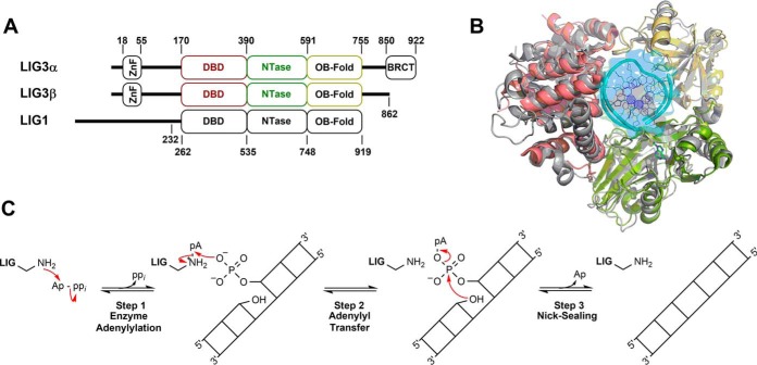
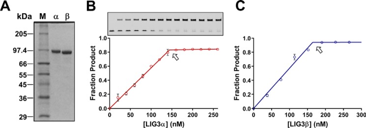
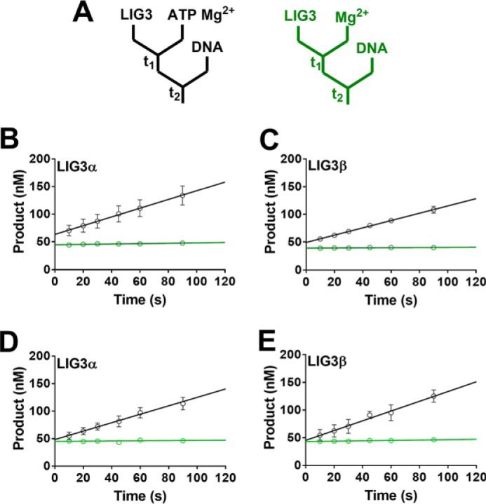
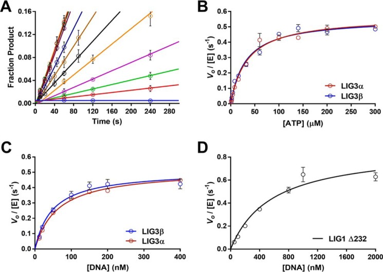
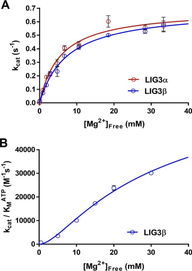
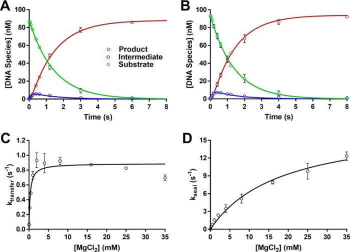
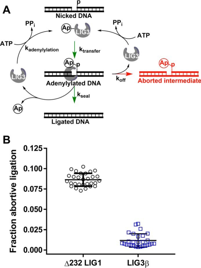
Humans have three genes encoding DNA ligases with conserved structural features and activities, but they also have notable differences. The LIG3 gene encodes a ubiquitous isoform in all tissues (LIG3α) and a germ line-specific splicing isoform (LIG3β) that differs in the C-terminal domain. Both isoforms are found in the nucleus and the mitochondria. Here, we determined the kinetics and thermodynamics of single-stranded break ligation by LIG3α and LIG3β and compared this framework to that of LIG1, the nuclear replicative ligase. The kinetic parameters of the LIG3 isoforms are nearly identical under all tested conditions, indicating that the BRCA1 C terminal (BRCT) domain specific to LIG3α does not alter ligation kinetics. Although LIG3 is only 22% identical to LIG1 across their conserved domains, the two enzymes had very similar maximal ligation rates. Comparison of the rate and equilibrium constants for LIG3 and LIG1 nevertheless revealed important differences. The LIG3 isoforms were seven times more efficient than LIG1 at ligating nicked DNA under optimal conditions, mainly because of their lower Km value for the DNA substrate. This could explain why LIG3 is less prone to abortive ligation than LIG1. Surprisingly, the affinity of LIG3 for Mg2+ was ten times weaker than that of LIG1, suggesting that Mg2+ availability regulates DNA ligation in vivo, because Mg2+ levels are higher in the mitochondria than in the nucleus. The biochemical differences between the LIG3 isoforms and LIG1 identified here will guide the understanding of both unique and overlapping biological roles of these critical enzymes.

Keywords: DNA ligase; DNA repair; DNA replication; abortive ligation; enzyme catalysis; magnesium; pre-steady-state kinetics; steady-state; steady-state kinetics.


Conflict of interest statement

The authors declare that they have no conflicts of interest with the contents of this article

Figures

Figure 1.
Figure 1.
Conserved structure and mechanism of human DNA ligases. A, schematic representation of human DNA LIG3α, LIG3β and LIG1. The numbers indicate domain boundaries of the enzymes. LIG3α contains a C-terminal BRCT domain. The LIG1 Δ232 truncation mutant used throughout this study is catalytically identical to full-length LIG1 (34). LIG3 isoforms and LIG1 possess conserved DBDs, NTase domains, and OB-fold domains. B, PyMOL (48) rendered superimposition of LIG1 (gray) and LIG3 (color-coded; DBD, red; NTase, green; OB-fold, yellow) structures with Protein Data Bank codes 1X9N and 3L2P, respectively (31, 32). C, mechanism of ATP-dependent DNA ligases.
Figure 2.
Figure 2.
Purity and active site titrations of LIG3 isoforms. A, 12% SDS-PAGE gel containing 0.5 μg of purified LIG3α and LIG3β with molecular masses of 102.7 and 95.9 kDa. M, protein sizing standards; α, Lig3α; β, Lig3β. B and C, representative active site titrations of LIG3α and LIG3β isoform, inset gel shows product (upper bands) and substrate (lower bands). In the absence of ATP, the adenylylated enzyme is limited to a single turnover ligation. Reactions contained 150 nm 28-mer nicked DNA substrate and 20 mm Mg2+ (see “Experimental procedures” for details). The equivalence point of the titration (denoted by arrow) indicates the concentration of adenylylated enzyme (data points are the means ± S.D.; n = 2).
Figure 3.
Figure 3.
Adenylylation state of purified LIG3 isoforms. A, schematics for burst experiments. The left (black) scheme indicates that LIG3 was incubated in the presence of ATP and Mg2+ to ensure complete enzyme adenylylation prior to the addition of DNA. The scheme to the right (green) displays reaction conditions in which no ATP was added to the reaction solution. The colors of the diagrams correspond to the colors of the linear fits in B–E. B and C, burst kinetics indicate LIG3α and LIG3β are 73 and 81% adenylylated, respectively. D and E, burst kinetics of LIG3α and LIG3β, respectively, after the addition of an adenylylation step during protein purification generates 100% adenylylated protein. Burst experiments after preincubation of LIG3 with ATP and Mg2+ did not increase burst amplitude. All experiments contained an estimated 50 nm enzyme, 200 nm DNA, and 20 mm Mg2+ in the presence and absence of 0.5 μm ATP. Each experiment was completed in triplicate (means ± S.D.).
Figure 4.
Figure 4.
Substrate dependence of LIG3α, LIG3β, and LIG1. A, representative ATP concentration dependence of LIG3β used to determine initial velocities under multiple-turnover conditions. Reactions performed at each ATP concentration (0–300 μm) were fit using linear regression to determine initial rates. B, ATP dependence was measured under multiple-turnover conditions in the presence of saturating DNA (1 μm) and Mg2+ (20 mm). Initial velocities are plotted as a function of ATP concentration and fit using the Michaelis–Menten equation yielding kcat, ATP values of 0.57 ± 0.02 and 0.55 ± 0.02 s−1 for LIG3α and LIG3β, respectively. The respective Km, ATP values are 34 ± 4 and 31 ± 3 μm. C, DNA dependence was measured at 300 mm ionic strength with 1 mm ATP and 20 mm Mg2+. LIG3α and LIG3β have kcat, DNA values of 0.52 ± 0.02 and 0.51 ± 0.03 s−1 and Km, DNA values of 62 ± 6 and 49 ± 8 nm, respectively. D, LIG1 DNA concentration dependence was measured under the same condition as for LIG3 in C. The kcat, DNA value for LIG1 is 0.87 ± 0.10 s−1 and the Km, DNA value is 570 ± 170 nm. Each experiment was completed in triplicate (means ± S.D.).
Figure 5.
Figure 5.
Magnesium dependence for multiple-turnover ligation. A, reactions contained 1 μm DNA, 1 mm ATP, and the concentration of free Mg2+ was varied between 0 and 35 mm. The data were fit using a hyperbolic one site-specific binding equation (Equation 2) yielding maximal kcat, Mg values of 0.69 ± 0.04 and 0.69 ± 0.03 s−1 and KMg values of 5.6 ± 0.9 and 7.4 ± 0.7 mm for LIG3α and LIG3β, respectively. B, investigation of the Mg2+ concentration dependence of enzyme adenylylation. The Mg2+ concentration dependence was performed using subsaturating ATP concentrations (supplemental Fig. S4). The magnesium concentration dependence of (kcat/Km)ATP for LIG3β was fit using a two metal random binding model (Equation 3). Each experiment was completed in at least triplicate (means ± S.D.).
Figure 6.
Figure 6.
Single-turnover ligation kinetics. A and B, representative single turnover kinetics of DNA LIG3α (A) and LIG3β (B) indicate the two enzymes behave similarly under single-turnover conditions at 20 mm Mg2+. The data were fit using a two-step irreversible mechanism using the program Berkeley Madonna. The rate constants for adenylyl transfer are 0.7 and 0.8 s−1 and for nick sealing are 8.7 and 8.9 s−1 for LIG3α and LIG3β, respectively. Experiments contained 100 nm fluorescein-labeled DNA, 600 nm enzyme, and 20 mm MgCl2 with no added ATP. C, Mg2+ concentration dependence for the adenylyl transfer step using LIG3β. D, Mg2+ concentration dependence of the nick-sealing step for LIG3β. The data were fit using a hyperbolic one site-specific binding equation (Equation 2) yielding ktransfer and kseal values of 0.89 ± 0.03 and 19 ± 6 s−1, respectively. The KMg values for adenylyl transfer and nick-sealing steps are 0.30 ± 0.06 and 18 ± 3 mm, respectively. Each experiment was completed in at least triplicate (means ± S.D.).
Figure 7.
Figure 7.
Accumulation of adenylylated DNA intermediate. A, abortive ligation mechanism observed under Mg2+-starved conditions. Adenylylated ligase interacts with nicked DNA substrates, generating adenylylated DNA intermediate. The partitioning between the nick-sealing steps and abortive ligation is represented with kseal and koff, respectively. Following successful or abortive ligation, ligase becomes adenylylated, preventing the enzyme from rebinding the adenylylated DNA intermediate and completing the nick-sealing step. B, propensity of DNA ligases for abortive ligation. Free Mg2+ concentrations were 70-fold below KMg for LIG1 and LIG3β in all experiments. The abortive ligation data represent multiple time points obtained from multiple-turnover time courses conducted in quadruplicate (see “Experimental procedures” for details). Error bars indicate S.D. from the mean.

References

    1. Bentley D., Selfridge J., Millar J. K., Samuel K., Hole N., Ansell J. D., and Melton D. W. (1996) DNA ligase I is required for fetal liver erythropoiesis but is not essential for mammalian cell viability. Nat. Genet. 13, 489–491 - PubMed
    1. Bentley D. J., Harrison C., Ketchen A. M., Redhead N. J., Samuel K., Waterfall M., Ansell J. D., and Melton D. W. (2002) DNA ligase I null mouse cells show normal DNA repair activity but altered DNA replication and reduced genome stability. J. Cell Sci. 115, 1551–1561 - PubMed
    1. Puebla-Osorio N., Lacey D. B., Alt F. W., and Zhu C. (2006) Early embryonic lethality due to targeted inactivation of DNA ligase III. Mol. Cell Biol. 26, 3935–3941 - PMC - PubMed
    1. Barnes D. E., Stamp G., Rosewell I., Denzel A., and Lindahl T. (1998) Targeted disruption of the gene encoding DNA ligase IV leads to lethality in embryonic mice. Curr. Biol. 8, 1395–1398 - PubMed
    1. Frank K. M., Sekiguchi J. M., Seidl K. J., Swat W., Rathbun G. A., Cheng H. L., Davidson L., Kangaloo L., and Alt F. W. (1998) Late embryonic lethality and impaired V(D)J recombination in mice lacking DNA ligase IV. Nature 396, 173–177 - PubMed

Publication types