

The interleukin-like epithelial-mesenchymal transition inducer ILEI exhibits a non-interleukin-like fold and is active as a domain-swapped dimer
- PMID: 28751379
- PMCID: PMC5602407
- DOI: 10.1074/jbc.M117.782904
The interleukin-like epithelial-mesenchymal transition inducer ILEI exhibits a non-interleukin-like fold and is active as a domain-swapped dimer
Abstract
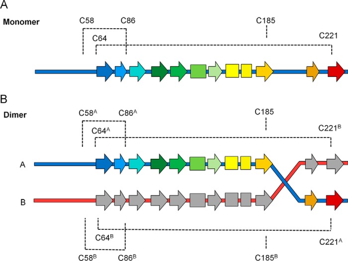
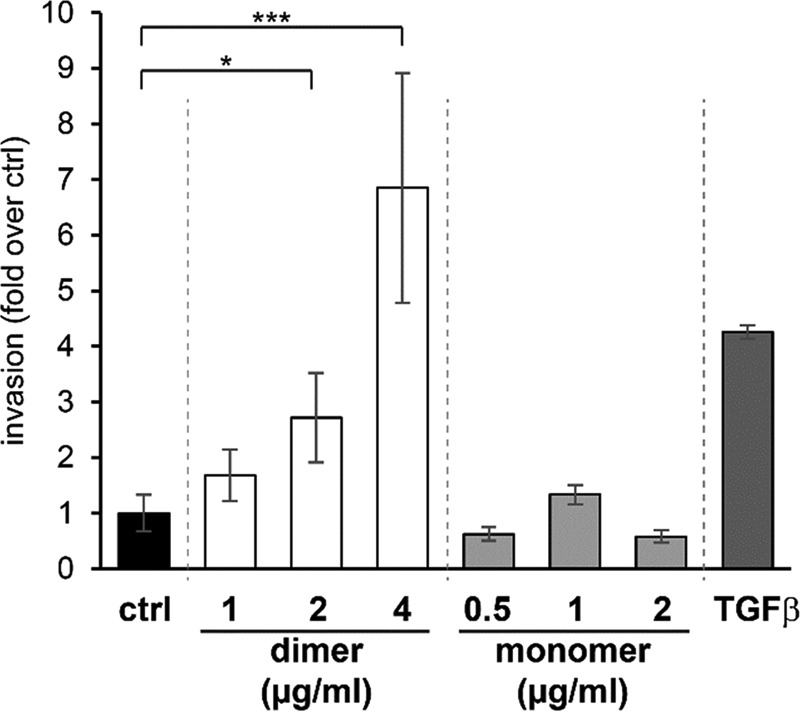
Production and secretion of pro-metastatic proteins is a feature of many tumor cells. The FAM3C interleukin-like epithelial-to-mesenchymal-transition (EMT) inducer (ILEI) has been shown to be strongly up-regulated in several cancers and to be essential for tumor formation and metastasis in epithelial cells, correlating with a significant decrease in overall survival in colon and breast cancer patients. ILEI has been seen to interact with the γ-secretase presenilin 1 subunit (PS1). However, not much is known about the mechanism-of-action or the detailed ILEI structure. We present here the crystal structures of FAM3C ILEI and show that it exists as monomers but also as covalent dimers. The observed ILEI β-β-α fold confirmed previous indications that the FAM3C proteins do not form classical four-helix-bundle structures as was initially predicted. This provides the first experimental evidence that the interleukin-like EMT inducers are not evolutionarily related to the interleukins. However, more surprisingly, the ILEI dimer structure was found to feature a trans-linked domain swap, converting an intramolecular disulfide to intermolecular. Interestingly, dimeric but not monomeric ILEI was subsequently found to cause a dose-dependent increase in EpRas cell invasiveness comparable with TGF-β, indicating that the dimer might be the active ILEI species. This is in line with a parallel study showing that covalent oligomerization of ILEI is essential for EMT and tumor progression in vivo The structures and the activity data give some first insight into the relationship between dimerization and ILEI function as well as indicate an intriguing link between ILEI, the PS1-protease, TGF-β, and the TGF-β receptor 1.
Keywords: cancer; crystallography; epithelial-mesenchymal transition (EMT); presenilin; transforming growth factor β (TGF-β); γ-secretase.
© 2017 by The American Society for Biochemistry and Molecular Biology, Inc.
Conflict of interest statement
A. M. J., J. M., A.-C. N., E. A., P. J., and L. H. S. are employees of AstraZeneca, which provided funding.
Figures

References
-
- Zhu Y., Xu G., Patel A., McLaughlin M. M., Silverman C., Knecht K., Sweitzer S., Li X., McDonnell P., Mirabile R., Zimmerman D., Boyce R., Tierney L. A., Hu E., Livi G. P., Wolf B., Abdel-Meguid S. S., Rose G. D., et al. (2002) Cloning, expression, and initial characterization of a novel cytokine-like gene family. Genomics 80, 144–150 - PubMed
-
- Waerner T., Alacakaptan M., Tamir I., Oberauer R., Gal A., Brabletz T., Schreiber M., Jechlinger M., and Beug H. (2006) ILEI: A cytokine essential for EMT, tumor formation, and late events in metastasis in epithelial cells. Cancer Cell 10, 227–239 - PubMed
-
- Csiszar A., Kutay B., Wirth S., Schmidt U., Macho-Maschler S., Schreiber M., Alacakaptan M., Vogel G. F., Aumayr K., Huber L. A., Beug H. (2014) Interleukin-like epithelial-to-mesenchymal transition inducer activity is controlled by proteolytic processing and plasminogen-urokinase plasminogen activator receptor system-regulated secretion during breast cancer progression. Breast Cancer Res. 16, 433. - PMC - PubMed
-
- Gao Z. H., Lu C., Wang Z. N., Song Y. X., Zhu J. L., Gao P., Sun J. X., Chen X. W., Wang M. X., Dong Y. L., and Xu H. M. (2014) ILEI: a novel target for Epithelial-Mesenchymal Transition and poor prognosis in colorectal cancer. Histopathology 65, 527–538 - PubMed
Publication types
MeSH terms
Substances
Associated data
- Actions
- Actions
- Actions
- Actions
- Actions
- Actions
Grants and funding
LinkOut - more resources
Full Text Sources
Other Literature Sources
Molecular Biology Databases
Miscellaneous
