. 2017 Oct;102(4):1093-1102.
doi: 10.1189/jlb.1A0417-147RR. Epub 2017 Jul 27.

Parkinson disease-associated LRRK2 G2019S transgene disrupts marrow myelopoiesis and peripheral Th17 response

Affiliations

Parkinson disease-associated LRRK2 G2019S transgene disrupts marrow myelopoiesis and peripheral Th17 response

Jeongho Park et al. J Leukoc Biol. 2017 Oct.

Abstract

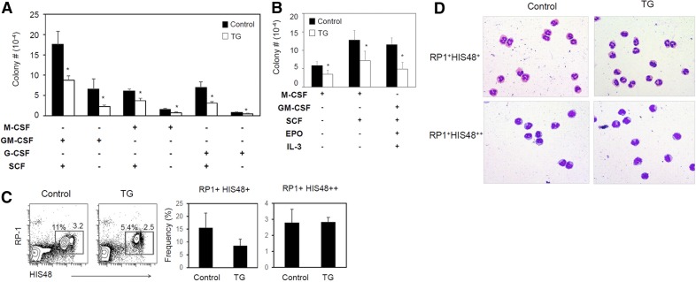
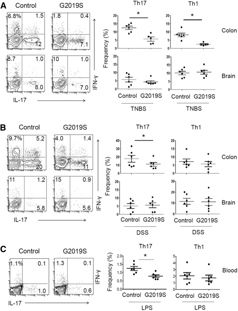
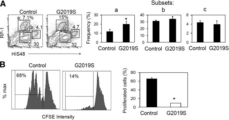
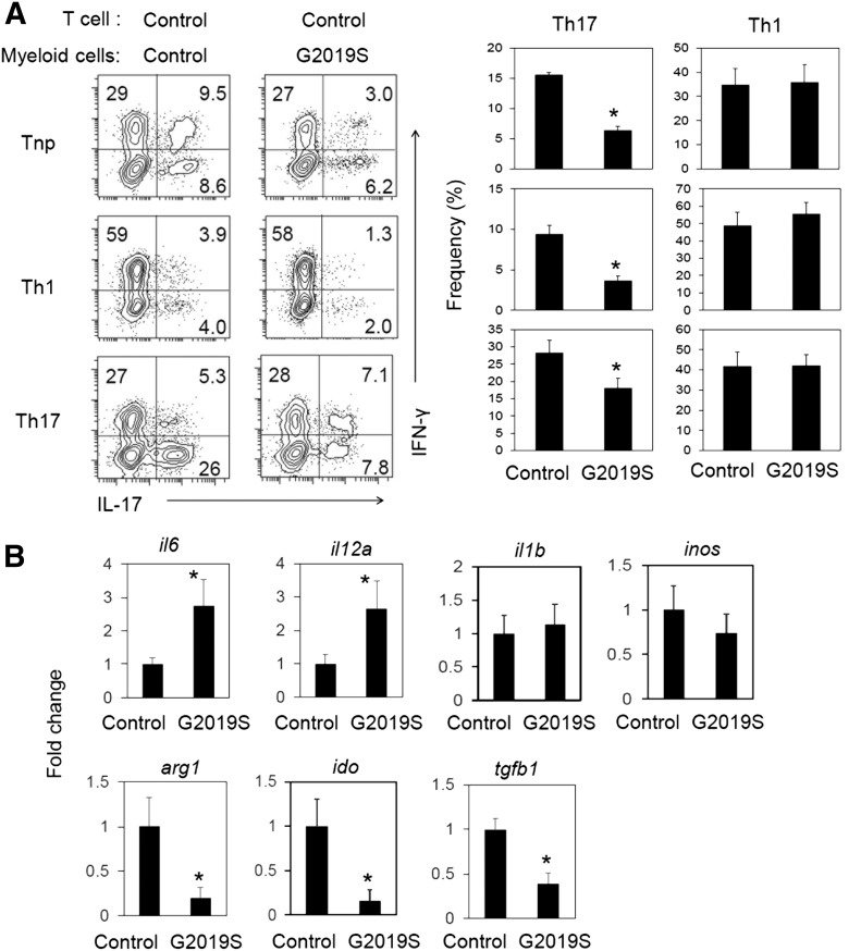
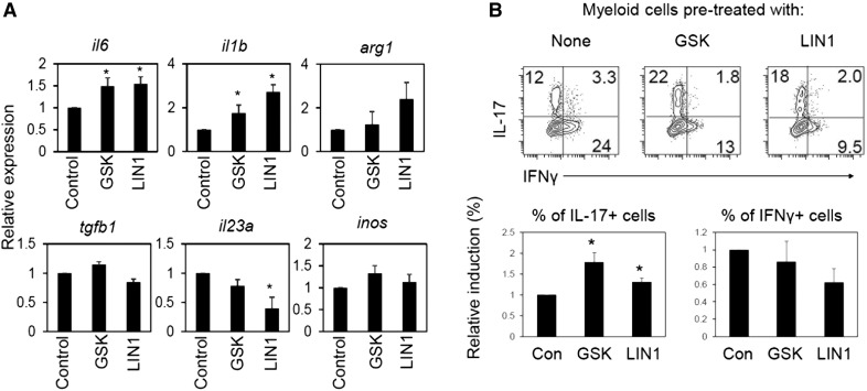
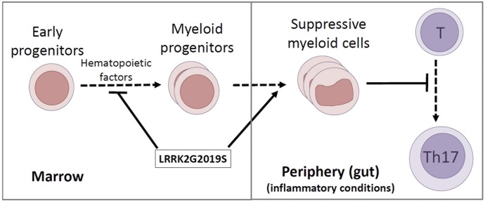
Parkinson's disease (PD) is a neurodegenerative disease, whereas Crohn's disease is an inflammatory bowel disease. Interestingly, polymorphisms in the LRRK2 gene have been identified as risk factors for both diseases. LRRK2 G2019S is the most prevalent mutation found in PD. To gain insights into the role of the LRRK2 G2019S gene on the development and activation of the immune system in the brain-gut axis, we investigated the effect of LRRK2 G2019S on bone marrow myeloid progenitors and myeloid cell function in the periphery. We used bacterial artificial chromosome transgenic rats harboring the human LRRK2 G2019S gene. LRRK2 G2019S transgene decreased the numbers of monocytic and granulocytic progenitors in the bone marrow. However, the numbers of peripheral, immature myeloid cells with suppressive activity were increased in the gut and blood circulation of LRRK2 G2019S compared with control rats in various acute and chronic inflammatory responses. In inflammatory conditions, Th17 cell activity was suppressed, but tissue-associated phylum Bacteroidetes was abnormally increased in the intestine of LRRK2 G2019S rats. The abnormally expanded myeloid cells because of the LRRK2 G2019S gene were highly suppressive on Th17 cell differentiation. Moreover, we found that inhibition of LRRK2 kinase affects myeloid progenitors and myeloid cell differentiation. Taken together, the results indicate that abnormal LRRK2 activity can alter bone marrow myelopoiesis, peripheral myeloid cell differentiation, and intestinal immune homeostasis. These findings may have ramifications in immune and inflammatory responses in patients with LRRK2 abnormalities.

Keywords: Crohn; T cells; inflammation; intestine; myeloid cells.


Figures

Figure 1.
Figure 1.. Decreased myelopoiesis in LRRK2 G2019S rats.
(A) Numbers of myeloid cell colonies formed in agar from the bone marrow progenitors of control and LRRK2 G2019S rats. (B) Numbers of myeloid cell colonies formed in methylcellulose from the bone marrow of control and LRRK2 G2019S rats. (C) Expression of RP-1 and HIS48 by peripheral blood myeloid cells. (D) Wright-Giemsa staining of sorted RP-1+ cell subsets. Blood RP-1+ cells were sorted into HIS48+ and HIS48++ cells. Representative dot plots and images and pooled data (n = 3–6) from 2–3 individual experiments are shown. *Significant differences (P ≤ 0.05) from control groups. Error bars indicate means ± sem.
Figure 2.
Figure 2.. Increased myeloid cell expansion in the intestine and blood, but not the brain, of LRRK2 G2019S rats challenged with TNBS, DSS, or LPS.
(A) RP-1+ cell frequency was examined in the colonic lamina propria. Control and LRRK2 G2019S TG rats were administered TNBS (rectal injection, 50 mg/kg of body weight) or DSS (4∼5%) as described in Supplemental Fig. 1. Rats were sacrificed 6 (TNBS) or 25 (DSS) d later, and indicated organs and tissues were examined for RP-1+ cell frequency. (B) Control and LRRK2 G2019S TG rats were injected with LPS (1.2 million EU/kg per on d 0) and sacrificed 15 d later, and blood cells were examined for RP-1+ cell frequency. (C) Colon tissues were examined for il17a mRNA expression. Expression levels were normalized by that of β-actin. (D) Colon tissues were examined for levels of Bacteroidetes based on Bacteroidetes-specific 16S rRNA gene levels. Tissue Bacteroidetes levels were normalized by β-actin gene levels. Representative dot plots and pooled data (n = 3–6) from 2 individual experiments are shown. *Significant differences (P ≤ 0.05) from control groups. Error bars indicate means ± sem.
Figure 3.
Figure 3.. Th17 cell frequency in the colon, brain, and blood of LRRK2 G2019S rats during intestinal or systemic inflammatory responses.
Frequencies of IL-17+ Th17 cells and IFN-γ+ Th1 cells in the colon, brain, or blood cells were examined in rats challenged with TNBS (A), DSS (B), and LPS (C). Control and G2019S rats were treated with TNBS, DSS, or LPS, as described in Supplemental Fig. 1. Rats were sacrificed 6 (A), 25 (B), or 15 d (C) later, and cells were isolated from the indicated tissues were examined for intracellular cytokine expression. Representative dot plots and pooled data (n = 5–6) from 2 individual experiments are shown. *Significant differences (P ≤ 0.05) from control groups. Error bars indicate means ± sem.
Figure 4.
Figure 4.. LRRK2 G2019S mutation promotes generation of suppressive myeloid cells in vitro.
(A) Rat bone marrow cells were differentiated with mM-CSF (20 ng/ml) for 7 d, and examined for RP-1 and HIS48. (B) T cell proliferation-suppression activity of myeloid cells was examined. Myeloid cells prepared as in (A) with M-CSF (20 ng/ml) for 7 d and then activated with LPS (1 μg/ml) for 24 h before coculture with isolated rat CD4+ T cells in the presence of SEB (5 μg/ml) for 3 d. Representative dot plots and pooled data (n = 3) are shown. *Significant differences (P ≤ 0.05) from control groups. Error bars indicate means ± sem.
Figure 5.
Figure 5.. LRRK2 G2019S gene in myeloid cells suppresses Th17 cell differentiation in vitro.
(A) Effects of in vitro–cultured myeloid cells on CD4+ T cell differentiation. LPS-activated myeloid cells were cocultured with rat spleen CD4+ T cells in the presence of SEB (5 μg/ml) for 5–6 d. Frequencies of IL-17+ Th17 cells and IFN-γ+ Th1 cells were examined after coculture. (B) Expression of selected genes in cultured myeloid cells. Rat bone marrow cells were differentiated with M-CSF (20 ng/ml) for 7 d and then activated with LPS for 24 h before quantitative RT-PCR analysis of indicated genes. Representative dot plots and pooled data (n = 5) are shown. *Significant differences (P ≤ 0.05) from control groups. Error bars indicate means ± sem.
Figure 6.
Figure 6.. Inhibition of LRRK2 kinase activity affects myeloid cell differentiation and phenotype.
(A) Effects of LRRK2 kinase inhibitors on selected gene expression in bone marrow–derived myeloid cells. (B) Function of bone marrow–derived myeloid cells, pretreated with LRRK2 kinase inhibitors, in regulating CD4+ T cell differentiation into Th17 and Th1 cells. Rat bone marrow cells were differentiated with M-CSF (20 ng/ml) for 7 d in the presence of indicated LRRK2 inhibitors (GSK2578215A or LRRK2-IN-1) and then activated with LPS for 24 h before quantitative RT-PCR analysis of indicated genes in (A) or for coculture in (B). LPS-activated myeloid cells were cocultured with rat spleen CD4+ T cells in the presence of SEB (5 μg/ml) for 5–6 d, and frequencies of IL-17+ Th17 cells and IFN-γ+ Th1 cells were examined after coculture. Representative dot plots and pooled data (n = 5) are shown. *Significant differences (P ≤ 0.05) from control groups. Error bars indicate means ± sem.
Figure 7.
Figure 7.. LRRK2 G2019S gene affects bone marrow myelopoiesis and peripheral myeloid cell differentiation and function.
The LRRK2 G2019S gene decreases the numbers of myeloid progenitors in the bone marrow of transgenic rats but abnormally increases peripheral expansion of suppressive myeloid cells, which suppress Th17 cell differentiation. Because myeloid cells and Th17 cells have central roles in promoting immunity and mediating inflammatory responses, the LRRK2 G2019S–induced dysregulation is likely to have significant effects on the inflammatory responses in various tissues.

Similar articles

Cited by

References

    1. Nichols W. C., Pankratz N., Hernandez D., Paisán-Ruíz C., Jain S., Halter C. A., Michaels V. E., Reed T., Rudolph A., Shults C. W., Singleton A., Foroud T.; Parkinson Study Group-PROGENI investigators (2005) Genetic screening for a single common LRRK2 mutation in familial Parkinson’s disease. Lancet 365, 410–412. - PubMed
    1. Di Fonzo A., Rohé C. F., Ferreira J., Chien H. F., Vacca L., Stocchi F., Guedes L., Fabrizio E., Manfredi M., Vanacore N., Goldwurm S., Breedveld G., Sampaio C., Meco G., Barbosa E., Oostra B. A., Bonifati V.; Italian Parkinson Genetics Network (2005) A frequent LRRK2 gene mutation associated with autosomal dominant Parkinson’s disease. Lancet 365, 412–415. - PubMed
    1. Gilks W. P., Abou-Sleiman P. M., Gandhi S., Jain S., Singleton A., Lees A. J., Shaw K., Bhatia K. P., Bonifati V., Quinn N. P., Lynch J., Healy D. G., Holton J. L., Revesz T., Wood N. W. (2005) A common LRRK2 mutation in idiopathic Parkinson’s disease. Lancet 365, 415–416. - PubMed
    1. Greggio E. (2012) Role of LRRK2 kinase activity in the pathogenesis of Parkinson’s disease. Biochem. Soc. Trans. 40, 1058–1062. - PubMed
    1. Rudenko I. N., Cookson M. R. (2014) Heterogeneity of leucine-rich repeat kinase 2 mutations: genetics, mechanisms and therapeutic implications. Neurotherapeutics 11, 738–750. - PMC - PubMed

Publication types

MeSH terms

Substances