

Sox10+ Cells Contribute to Vascular Development in Multiple Organs-Brief Report
- PMID: 28751573
- PMCID: PMC5572822
- DOI: 10.1161/ATVBAHA.117.309774
Sox10+ Cells Contribute to Vascular Development in Multiple Organs-Brief Report
Abstract
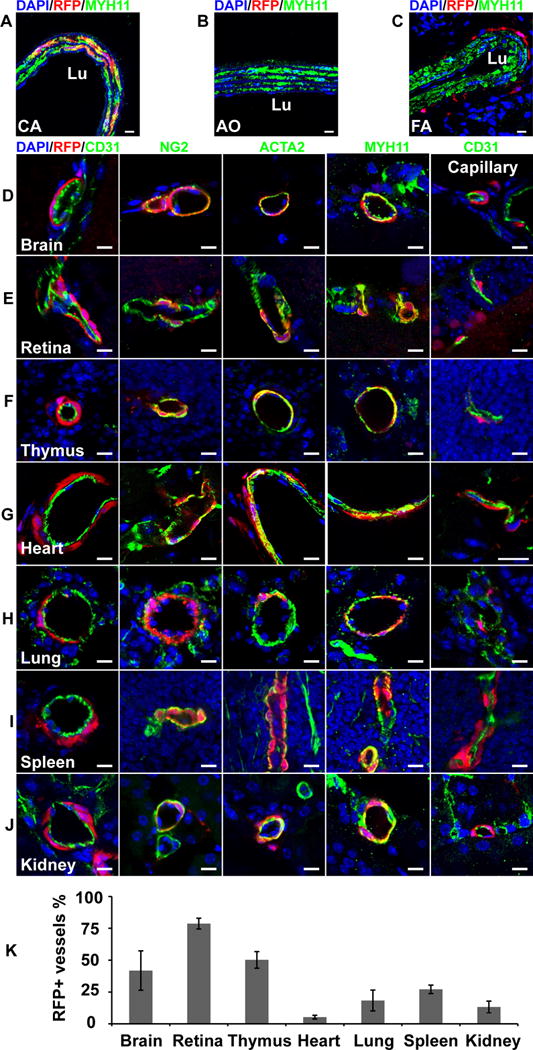
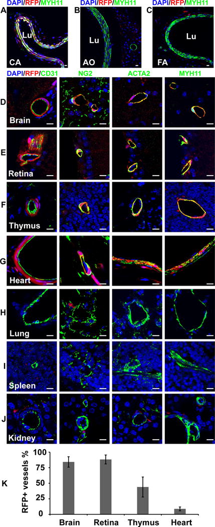
Objective: Previous genetic lineage tracing studies showed that Sox10+ cells differentiate into vascular mural cells, limited to neural crest-derived blood vessels in craniofacial tissues, aortic arch, pulmonary arch arteries, brachiocephalic, carotid arteries, and thymus. The purpose of this study was to investigate the contribution of Sox10+ cells to the vascular development in other tissues and organs and their relationship with neural crest.
Approach and results: Using genetic lineage tracing technique based on Cre/LoxP system, we examined blood vessels in the adult organs of the mice expressing Sox10-Cre/Rosa-LoxP-red fluorescent protein or Wnt1-Cre/Rosa-LoxP-red fluorescent protein by immunohistological analysis. In addition to previously reported tissues and organs derived from neural crest, we showed that Sox10+ cells also contributed to vascular mural cells in the lung, spleen, and kidney, which are derived from non-neural crest origin as evidenced by red fluorescent protein-negative blood vessels in these 3 organs of Wnt1-Cre/Rosa-LoxP-red fluorescent protein mice.
Conclusions: This study demonstrates that Sox10+ cells contribute to pericytes and smooth muscle cells in most parts of the body, including those from neural crest and non-neural crest, which has significant implications in vascular remodeling under physiological and pathological conditions.
Keywords: blood vessel; mouse; pericyte; smooth muscle cell; vascular remodeling.
© 2017 American Heart Association, Inc.
Figures
References
-
- Gittenberger-de Groot AC, DeRuiter MC, Bergwerff M, Poelmann RE. Smooth muscle cell origin and its relation to heterogeneity in development and disease. Arterioscler Thromb Vasc Biol. 1999;19:1589–1594. - PubMed
-
- Majesky MW. Developmental basis of vascular smooth muscle diversity. Arterioscler Thromb Vasc Biol. 2007;27:1248–1258. - PubMed
-
- Bergwerff M, Verberne ME, DeRuiter MC, Poelmann RE, Gittenberger-de Groot AC. Neural crest cell contribution to the developing circulatory system: Implications for vascular morphology? Circ Res. 1998;82:221–231. - PubMed
-
- Etchevers HC, Vincent C, Le Douarin NM, Couly GF. The cephalic neural crest provides pericytes and smooth muscle cells to all blood vessels of the face and forebrain. Development. 2001;128:1059–1068. - PubMed
-
- Korn J, Christ B, Kurz H. Neuroectodermal origin of brain pericytes and vascular smooth muscle cells. J Comp Neurol. 2002;442:78–88. - PubMed
Publication types
MeSH terms
Substances
Grants and funding
LinkOut - more resources
Full Text Sources
Other Literature Sources
Molecular Biology Databases
