

Expression of ADAMTS13 in Normal and Abnormal Placentae and Its Potential Role in Angiogenesis and Placenta Development
- PMID: 28751574
- PMCID: PMC5570641
- DOI: 10.1161/ATVBAHA.117.309735
Expression of ADAMTS13 in Normal and Abnormal Placentae and Its Potential Role in Angiogenesis and Placenta Development
Abstract
Objective: ADAMTS13 (a disintegrin and metalloproteinase with thrombospondin type 1 repeats, member 13) is primarily synthesized in liver. The biosynthesis of ADAMTS13 and its physiological role in placenta are not known.
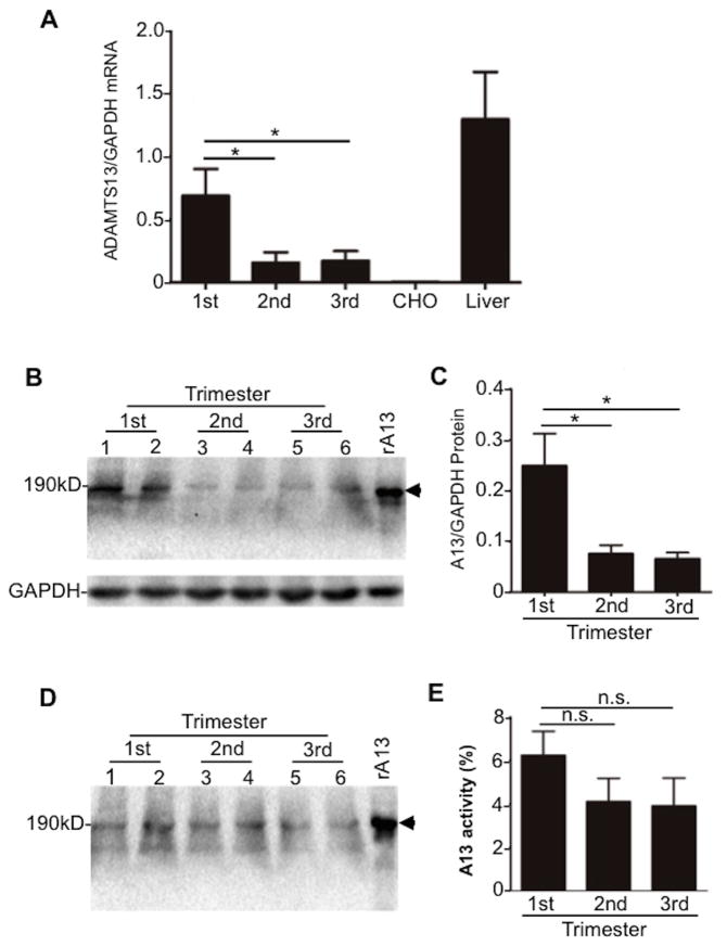
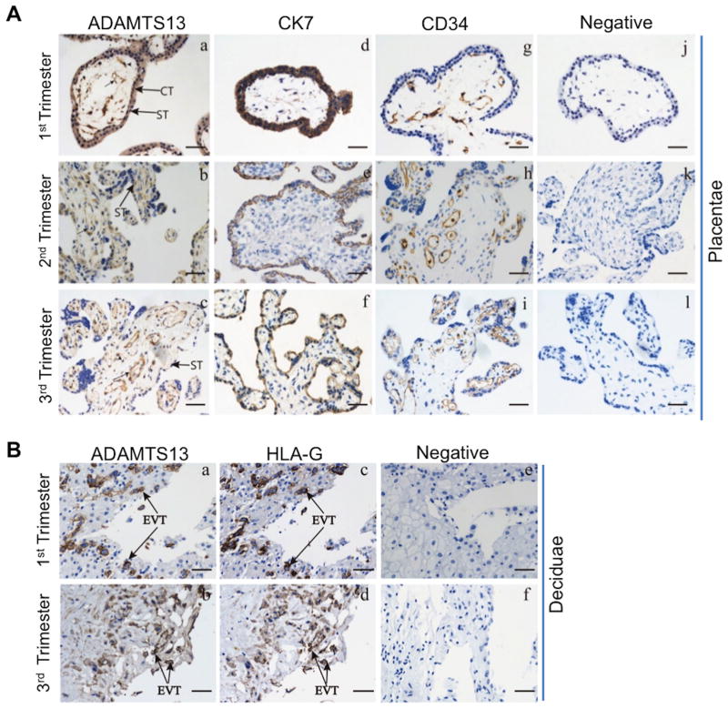
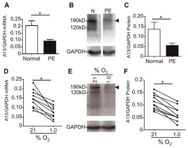
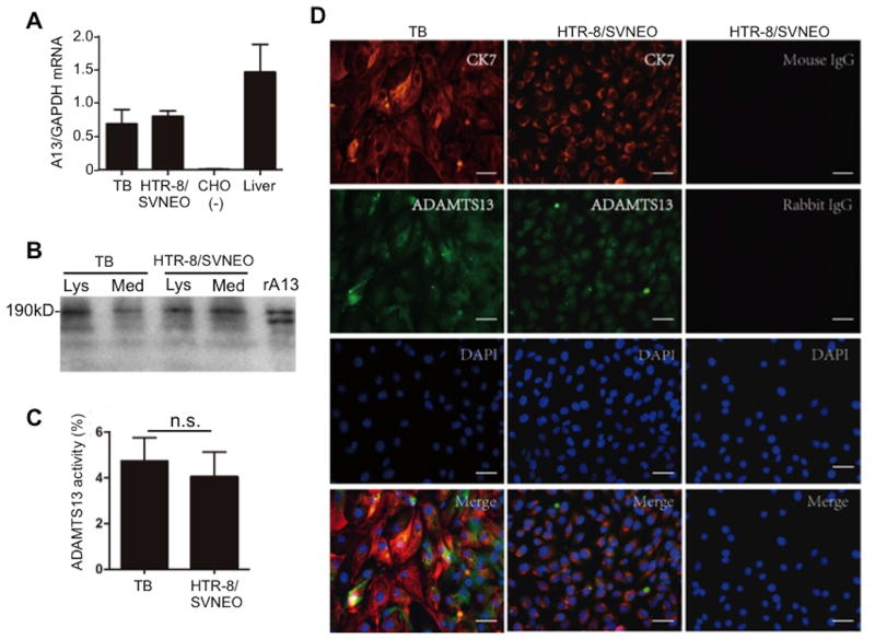
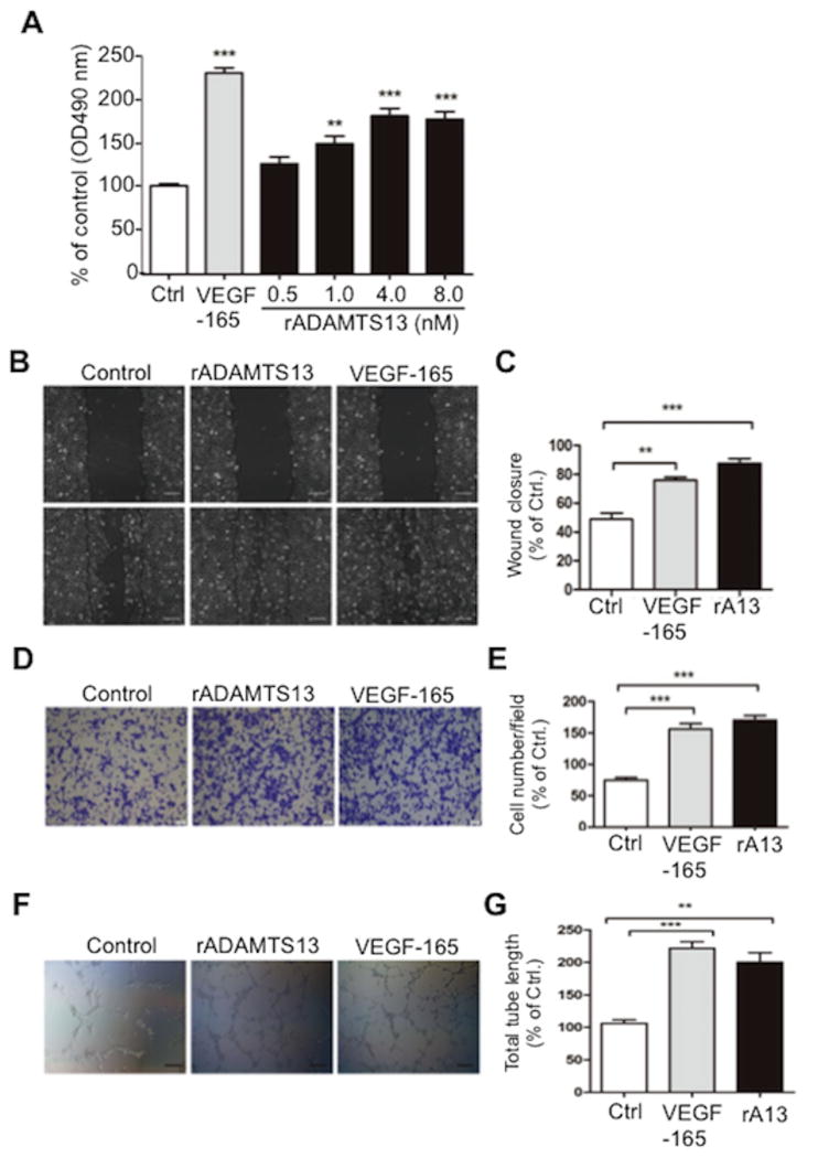
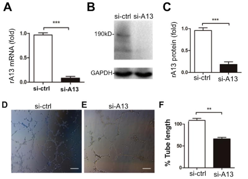
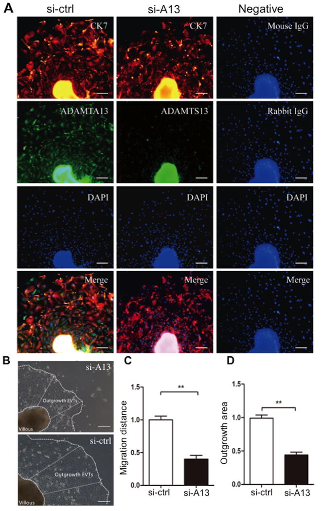
Approach and results: We used real-time polymerase chain reaction, immunohistochemistry, and Western blotting analyses, as well as proteolytic cleavage of FRETS (fluorescent resonance energy transfers)-VWF73, to determine ADAMTS13 expression in placenta and trophoblasts obtained from individuals with normal pregnancy and patients with severe preeclampsia. We also determined the role of ADAMTS13 in extravillous trophoblasts using a 3-(4,5-dimethylthiazol-2-yl)-2,5-diphenyltetrazolium bromide assay, wound scratch assay, transwell migration assay, tube formation assay, and tissue outgrowth assays. We showed that full-length and proteolytically active ADAMTS13 was expressed in normal human placenta, primarily in the trophoblasts and villous core fetal vessel endothelium during pregnancy. Placental expression of ADAMTS13 mRNA, protein, and proteolytic activity was at the highest levels during the first trimester and significantly reduced at the term of gestation. Additionally, significantly reduced levels of placental ADAMTS13 expression was detected under hypoxic conditions and in patients with preeclampsia. In addition, recombinant ADAMTS13 protease stimulated proliferation, migration, invasion, and network formation of trophoblastic cells in culture. Finally, knockdown of ADAMTS13 expression attenuated the ability of tube formation in trophoblast (HTR-8/SVNEO) cells and the extravillous trophoblast outgrowth in placental explants.
Conclusions: Our results demonstrate for the first time the expression of ADAMTS13 mRNA and protein in normal and abnormal placental tissues and its role in promoting angiogenesis and trophoblastic cell development. The findings support the potential role of the ADAMTS13-von Willebrand factor pathway in normal pregnancy and pathogenesis of preeclampsia.
Keywords: ADAMTS13; placenta; preeclampsia; protein synthesis; trophoblast.
© 2017 American Heart Association, Inc.
Conflict of interest statement
Figures
Similar articles
-
miR-15b-AGO2 play a critical role in HTR8/SVneo invasion and in a model of angiogenesis defects related to inflammation.Placenta. 2016 May;41:62-73. doi: 10.1016/j.placenta.2016.03.007. Epub 2016 Mar 14. Placenta. 2016. PMID: 27208409
-
The chemokine CXCL6 restricts human trophoblast cell migration and invasion by suppressing MMP-2 activity in the first trimester.Hum Reprod. 2013 Sep;28(9):2350-62. doi: 10.1093/humrep/det258. Epub 2013 Jun 28. Hum Reprod. 2013. PMID: 23814098
-
Expression of Gadd45α in human early placenta and its role in trophoblast invasion.Placenta. 2014 Jun;35(6):370-7. doi: 10.1016/j.placenta.2014.03.020. Epub 2014 Apr 12. Placenta. 2014. PMID: 24755561
-
PPARgamma and early human placental development.Curr Med Chem. 2008;15(28):3011-24. doi: 10.2174/092986708786848677. Curr Med Chem. 2008. PMID: 19075649 Review.
-
Why is placentation abnormal in preeclampsia?Am J Obstet Gynecol. 2015 Oct;213(4 Suppl):S115-22. doi: 10.1016/j.ajog.2015.08.042. Am J Obstet Gynecol. 2015. PMID: 26428489 Free PMC article. Review.
Cited by
-
ADAMTS13 regulates angiogenic markers via Ephrin/Eph signaling in human mesenchymal stem cells under serum-deprivation stress.Sci Rep. 2024 Jan 4;14(1):560. doi: 10.1038/s41598-023-51079-z. Sci Rep. 2024. PMID: 38177376 Free PMC article.
-
Thrombotic Microangiopathy in Pregnancy: Current Understanding and Management Strategies.Kidney Int Rep. 2024 May 22;9(8):2353-2371. doi: 10.1016/j.ekir.2024.05.016. eCollection 2024 Aug. Kidney Int Rep. 2024. PMID: 39156177 Free PMC article. Review.
-
[Relationship Between ABO Blood Group and Pregnancy Complications].Sichuan Da Xue Xue Bao Yi Xue Ban. 2022 Sep;53(5):935-940. doi: 10.12182/20220960304. Sichuan Da Xue Xue Bao Yi Xue Ban. 2022. PMID: 36224700 Free PMC article. Review. Chinese.
-
Increased Complement Activation and Decreased ADAMTS13 Activity Are Associated with Genetic Susceptibility in Patients with Preeclampsia/HELLP Syndrome Compared to Healthy Pregnancies: An Observational Case-Controlled Study.J Pers Med. 2024 Apr 3;14(4):387. doi: 10.3390/jpm14040387. J Pers Med. 2024. PMID: 38673014 Free PMC article.
-
Reporting Sex and Sex Differences in Preclinical Studies.Arterioscler Thromb Vasc Biol. 2018 Oct;38(10):e171-e184. doi: 10.1161/ATVBAHA.118.311717. Arterioscler Thromb Vasc Biol. 2018. PMID: 30354222 Free PMC article. No abstract available.
References
-
- Hypertension in pregnancy. Report of the American College of Obstetricians and Gynecologists' Task Force on Hypertension in Pregnancy. Obstet Gynecol. 2013;122:1122–1131. - PubMed
-
- Herzog EM, Eggink AJ, Reijnierse A, Kerkhof MA, de Krijger RR, Roks AJ, Reiss IK, Nigg AL, Eilers PH, Steegers EA, Steegers-Theunissen RP. Impact of early- and late-onset preeclampsia on features of placental and newborn vascular health. Placenta. 2017;49:72–79. - PubMed
-
- Pauli JM, Repke JT. Preeclampsia: Short-term and Long-term Implications. Obstet Gynecol Clin North Am. 2015;42:299–313. - PubMed
-
- Staff AC, Benton SJ, von Dadelszen P, Roberts JM, Taylor RN, Powers RW, Charnock-Jones DS, Redman CW. Redefining preeclampsia using placenta-derived biomarkers. Hypertension. 2013;61:932–942. - PubMed
Publication types
MeSH terms
Substances
Grants and funding
LinkOut - more resources
Full Text Sources
Other Literature Sources
