

ChromEMT: Visualizing 3D chromatin structure and compaction in interphase and mitotic cells
- PMID: 28751582
- PMCID: PMC5646685
- DOI: 10.1126/science.aag0025
ChromEMT: Visualizing 3D chromatin structure and compaction in interphase and mitotic cells
Abstract
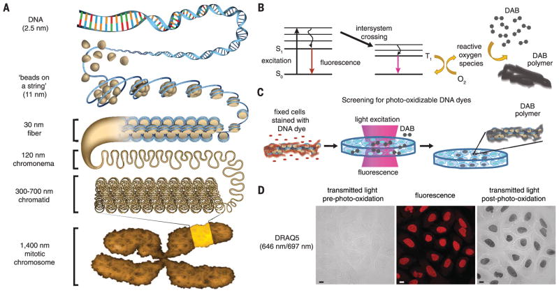
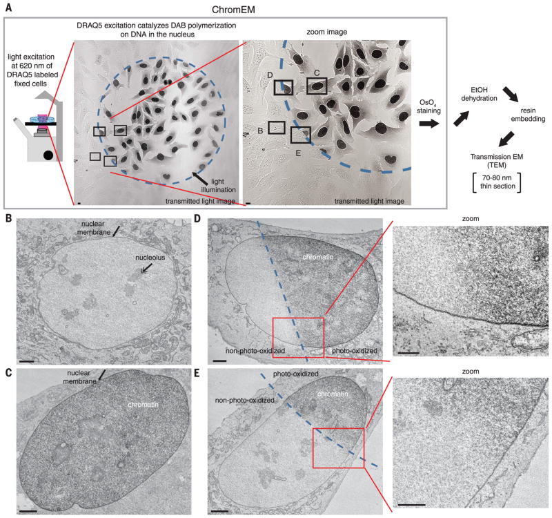
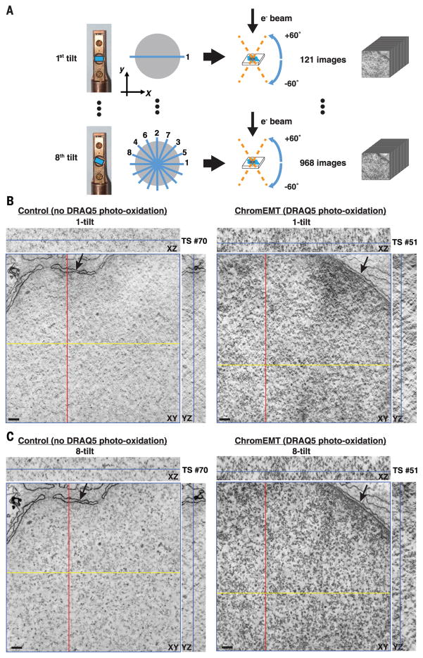
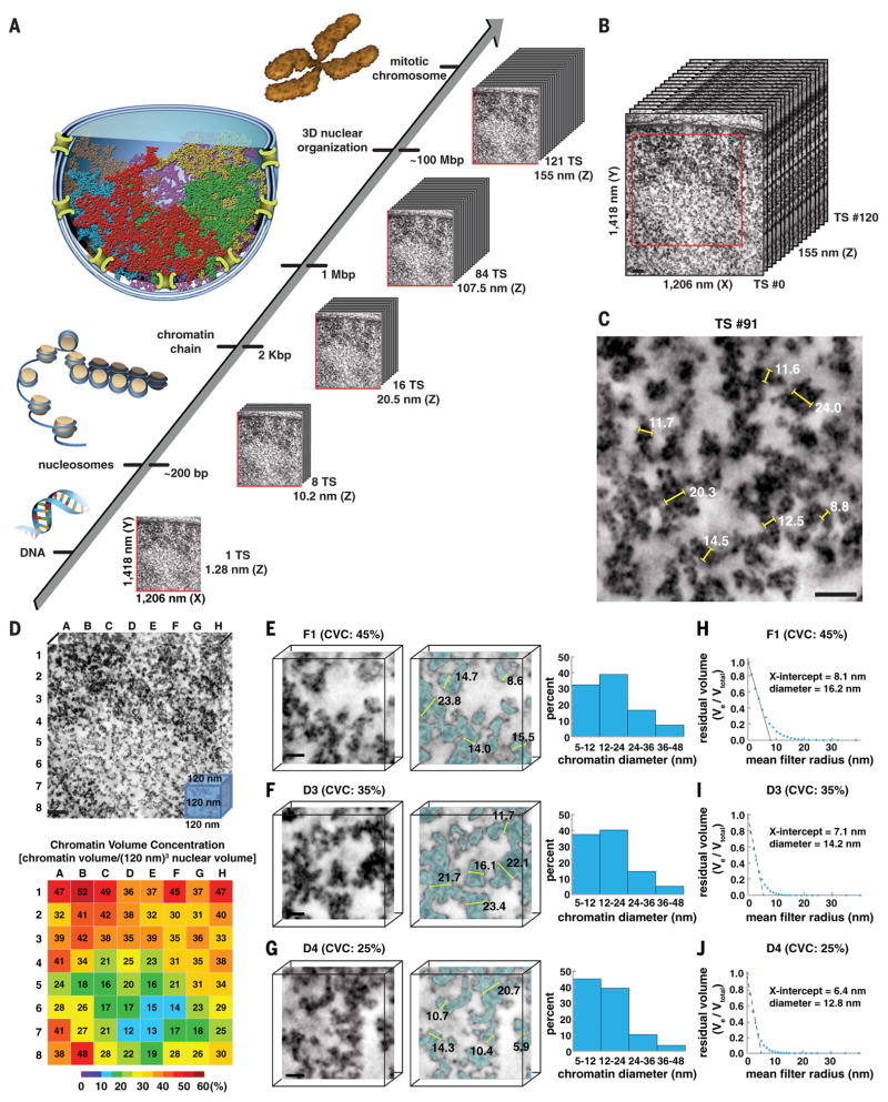
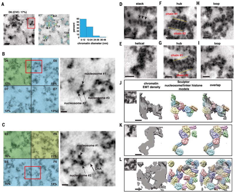
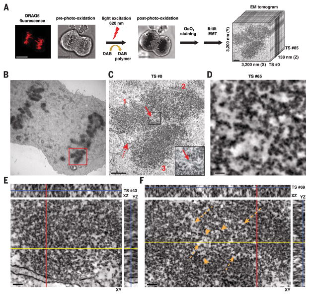
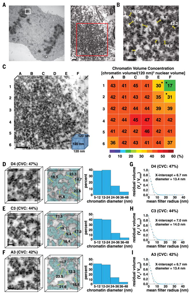
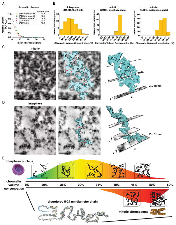
The chromatin structure of DNA determines genome compaction and activity in the nucleus. On the basis of in vitro structures and electron microscopy (EM) studies, the hierarchical model is that 11-nanometer DNA-nucleosome polymers fold into 30- and subsequently into 120- and 300- to 700-nanometer fibers and mitotic chromosomes. To visualize chromatin in situ, we identified a fluorescent dye that stains DNA with an osmiophilic polymer and selectively enhances its contrast in EM. Using ChromEMT (ChromEM tomography), we reveal the ultrastructure and three-dimensional (3D) organization of individual chromatin polymers, megabase domains, and mitotic chromosomes. We show that chromatin is a disordered 5- to 24-nanometer-diameter curvilinear chain that is packed together at different 3D concentration distributions in interphase and mitosis. Chromatin chains have many different particle arrangements and bend at various lengths to achieve structural compaction and high packing densities.
Copyright © 2017 The Authors, some rights reserved; exclusive licensee American Association for the Advancement of Science. No claim to original U.S. Government Works.
Figures


Comment in
-
Cover stories: A method for revealing chromatin.Science. 2017 Jul 28;357(6349):eaao4487. doi: 10.1126/science.aao4487. Science. 2017. PMID: 28751583 No abstract available.
-
The genome-seeing it clearly now.Science. 2017 Jul 28;357(6349):354-355. doi: 10.1126/science.aao1893. Science. 2017. PMID: 28751596 Free PMC article. No abstract available.
-
Genome organization: A vision of 3D chromatin organization.Nat Rev Mol Cell Biol. 2017 Aug 23;18(9):532. doi: 10.1038/nrm.2017.88. Nat Rev Mol Cell Biol. 2017. PMID: 28831176 No abstract available.
