. 2017 Jul 28;357(6349):378-381.
doi: 10.1126/science.aah6362.

Tough adhesives for diverse wet surfaces

Affiliations

Tough adhesives for diverse wet surfaces

J Li et al. Science. .

Abstract

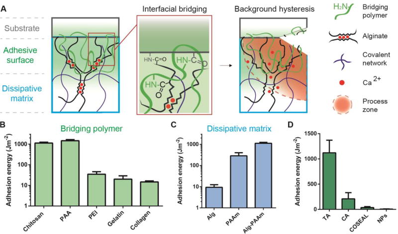
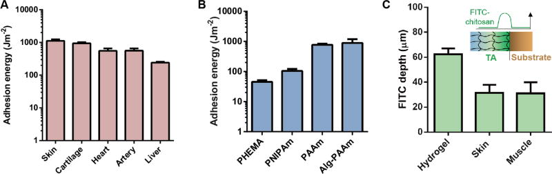
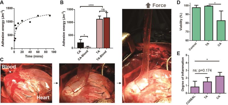
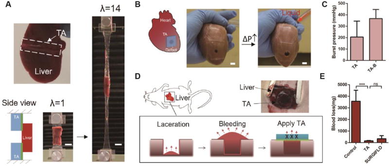
Adhesion to wet and dynamic surfaces, including biological tissues, is important in many fields but has proven to be extremely challenging. Existing adhesives are cytotoxic, adhere weakly to tissues, or cannot be used in wet environments. We report a bioinspired design for adhesives consisting of two layers: an adhesive surface and a dissipative matrix. The former adheres to the substrate by electrostatic interactions, covalent bonds, and physical interpenetration. The latter amplifies energy dissipation through hysteresis. The two layers synergistically lead to higher adhesion energies on wet surfaces as compared with those of existing adhesives. Adhesion occurs within minutes, independent of blood exposure and compatible with in vivo dynamic movements. This family of adhesives may be useful in many areas of application, including tissue adhesives, wound dressings, and tissue repair.


Figures

Fig. 1
Fig. 1. Design of tough adhesive (TA)
(A) TA consists of a dissipative matrix (light blue square) made of a hydrogel containing both ionically (calcium) crosslinked and covalently crosslinked polymers (black and blue lines), and an adhesive surface that contains a bridging polymer with primary amines (green lines). The bridging polymer penetrates into TA and the substrate (light green region). When a crack approaches, a process zone (orange area) dissipates significant energy as ionic bonds between alginate chains and calcium ions break. (B) Adhesion energy on porcine skin was measured using different bridging polymers. (C) Adhesion energy varies with the hydrogel matrix. (D) Comparison between TA and other adhesives. Error bars show standard deviation; N=4.
Fig. 2
Fig. 2. Adherence on diverse wet surfaces
TA adheres to a variety of (A) tissue surfaces and (B) hydrogels, including poly(hydroxyethyl methacrylate) (PHEMA), poly(N’-isopropylacrylamide) (PNIPAm), polyacrylamide (PAAm) and alginate-polyacrylamide (Alg-PAAm) hydrogels. (C) Penetration depth of FITC-chitosan into PAAm hydrogels, skin and muscle. Error bars show standard deviation; N=4.
Fig. 3
Fig. 3. Adhesion performance and biocompatibility
(A) Adhesion kinetics of TA to porcine skin. (B) Comparison of TA to cyanoacrylate (CA) placed on porcine skin with and without exposure to blood. N=4–6. (C) In vivo test on a beating porcine heart with blood exposure. (D) In vitro cell compatibility was compared by quantifying the viability of human dermal fibroblasts. N=4. (E) In vivo biocompatibility was evaluated using subcutaneous implantation in rats. Degree of inflammation was determined by a pathologist (0=normal, 1=very mild, 2=mild, 3=moderate, 4=severe, 5=very severe). Error bars show standard deviation; N=4–6. P values were determined by the t-test; *, p≤0.05; ****, p≤0.0001; ns, not significant.
Fig. 4
Fig. 4. Application enabled by TA
(A) TA was used as tissue adhesives. TA adhered to the liver and sustained 14 times its initial length before debonding. Scale bar, 20mm. (B) TA served as heart sealants. The TA sealant prevented liquid (red) leakage as the porcine heart was inflated. Scale bar, 10mm. (C) Burst pressures of the TA sealant were measured without and with plastic backing (TA-B). (D) Use of TA as hemostatic dressing. A deep wound was created on liver in rats, and then sealed with TA to stop the blood flow (labelled with red arrows). (E) Blood loss with the treatment of TA, SURGIFLO hemostat and control without treatment. Error bars show standard deviation; sample size N=4. P values were determined by the t-test; ***, p≤0.001; ns, not significant.

References

    1. Duflo S, Thibeault SL, Li W, Shu XZ, Prestwich GD. Vocal fold tissue repair in vivo using a synthetic extracellular matrix. Tissue Eng. 2006;12:2171–2180. - PubMed
    1. Sharma B, et al. Human cartilage repair with a photoreactive adhesive-hydrogel composite. Sci. Transl. Med. 2013;5:167ra6. - PMC - PubMed
    1. Prausnitz MR, Langer R. Transdermal drug delivery. Nat. Biotechnol. 2008;26:1261–1268. - PMC - PubMed
    1. Li J, Mooney DJ. Designing hydrogels for controlled drug delivery. Nat. Rev. Mater. 2016;1:16071. - PMC - PubMed
    1. Ghobril C, Charoen K, Rodriguez EK, Nazarian A, Grinstaff MW. A dendritic thioester hydrogel based on thiol–thioester exchange as a dissolvable sealant system for wound closure. Angew. Chem. Int. Ed. 2013;52:14070–14074. - PMC - PubMed

Publication types