. 2017 Jul 27;7(1):6690.
doi: 10.1038/s41598-017-06982-7.

Gene-based Therapy in a Mouse Model of Blue Cone Monochromacy

Affiliations

Gene-based Therapy in a Mouse Model of Blue Cone Monochromacy

Yuxin Zhang et al. Sci Rep. .

Erratum in

Abstract

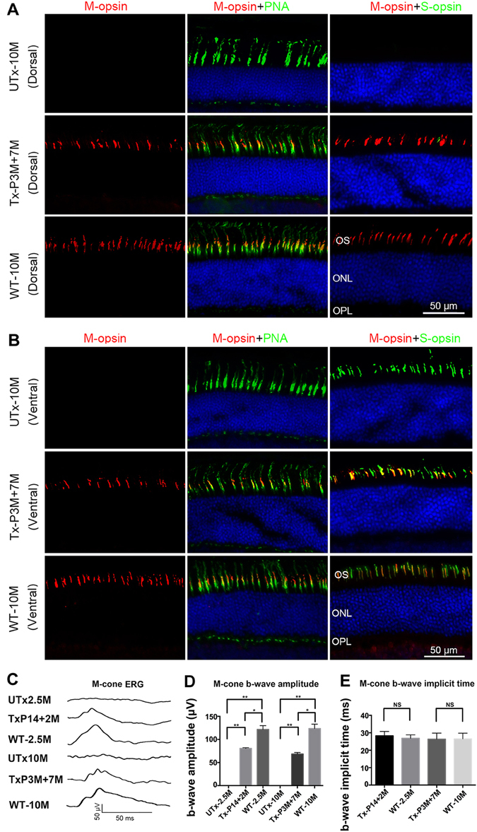
Cones are responsible for daylight, central, high acuity and color vision. Three proteins found in human cones, i.e. long-wavelength (L)-, middle-wavelength (M)-, and short-wavelength sensitive (S)-opsins, are responsible for red, green and blue color recognition, respectively. Human blue cone monochromacy (BCM) is characterized by functional loss of both L- and M-cone opsins due to mutations in the OPN1LW/OPN1MW gene cluster on the X chromosome. BCM patients, who rely on their vision from only S-cones and rods, suffer severely reduced visual acuity and impaired color vision. Recent studies show that there is sufficient cone structure remaining in the central fovea of BCM patients to consider AAV-mediated gene augmentation therapy. In contrast, mouse retina has only two opsins, S-opsin and M-opsin, but no L-opsin. We generated an M-opsin knockout mouse (Opn1mw -/-) expressing only S-opsin as a model for human BCM. We show that recombinant M-opsin delivered by AAV5 vectors rescues M-cone function in Opn1mw -/- mice. We also show that AAV delivered M-opsin localizes in the dorsal cone outer segments, and co-localizes with S-opsin in the ventral retina. Our study demonstrates that cones without M-opsin remain viable and respond to gene augmentation therapy, thereby providing proof-of-concept for cone function restoration in BCM patients.


Conflict of interest statement

WWH and the University of Florida have a financial interest in the use of AAV therapies; WWH owns equity in and is a consultant for AGTC Inc. that might, in the future, commercialize some aspects of this work.

Figures

Figure 1
Figure 1
Comparison of M- and S-opsin in Opn1mw−/− and WT mice. P14 Opn1mw−/− mice were treated with AAV5-PR2.1-mouse M-opsin vector and western blot analysis of mouse M-opsin (A) and S-opsin (B) was performed at 2 month post-injection with age-matched wild type eyes (WT); Tubulin was served as loading control. Diagram of M- and S-opsin distribution in WT retina was shown in (C). Frozen sections were prepared for immunostaining from dorsal (D) and ventral (E) retina of 2.5-month-old untreated Opn1mw−/− mice (UTx-2.5M), Opn1mw−/− mice treated at P14 and analyzed at 2 months post-injection (Tx-P14 + 2 M), and age-matched wild type control mice (WT-2.5M). (D) Analysis of the dorsal retina with M- and S-opsin antibodies and PNA. Neither M- nor S-opsin was detected in the dorsal retinas of the untreated eye (UTx-2.5M), but PNA staining was normal. In the treated eye (Tx-P14 + 2 M), AAV-delivered M-opsin was now abundant in the dorsal retina and co-localized with PNA. In an untreated age-matched wild type eye (WT-2.5M), M-opsin is found co-localized with PNA with very little or no S-opsin in the dorsal retina. (E) Analysis of the ventral retina. S-opsin expression and PNA staining were normal in the untreated Opn1mw−/− ventral retina (UTx-2.5M). AAV-delivered M-opsin co-localized with S-opsin (high magnification image is highlighted with a box) and PNA in cone outer segments in the treated Opn1mw−/− ventral retina (Tx-P14 + 2 M). In the untreated wild type ventral retina (UTx-2.5), M-opsin is expressed in the ventral retina at a lower level compared to that in the dorsal retina but is detectable and is co-localized with PNA and S-opsin. 50μm in each figure.
Figure 2
Figure 2
ERG analysis of middle wavelength cone function in mouse M-opsin treated Opn1mw−/− eyes and immunostaining of retinal sections from 10-month-old untreated Opn1mw−/− mice (UTx-10M), Opn1mw−/− mice treated at 3 months of age and analyzed 7 months post-injection (Tx-P3M + 7M), and age-matched wild type uninjected control mice (WT-10M). (A) Analysis of dorsal retinas. M- and S-opsin were not detected in the dorsal retinas in the untreated eye (UTx-10M), whereas PNA staining is normal. AAV-delivered M-opsin was co-localized with PNA in the treated dorsal retina (Tx-P3M + 7M). An age matched wild type retina (WT-10M) showed normal M-opsin expression and undetectable S-opsin expression in the dorsal retina. (B) Analysis of ventral retinas. S-opsin expression and PNA staining were normal in the untreated Opn1mw−/− ventral retina (UTx-10M). AAV-delivered M-opsin was co-localized with S-opsin and PNA in the treated Opn1mw−/− ventral retina (Tx-P3M + 7M). An age-matched wild type eye (WT-10M) showed normal S-opsin expression and a detectable level of M-opsin expression in the ventral retina. (C) Representative middle wavelength ERG recordings from UTx-2.5M: an untreated 2.5 month old Opn1mw−/− eye, TxP14 + 2 M: an Opn1mw−/− eye treated at P14 with ERG analysis at 2 months post-treatment, UTx-10M: an uninjected wild type eye at 10 months of age, TxP3M + 7 M: an Opn1mw−/− eye treated at 3 months of age with ERG analysis at 7 months post-treatment, and WT-10M: an uninjected wild type eye at 10 months of age. (D) Average b-wave amplitudes of middle wavelength ERGs from the above mice (n = 4 in each group), **P < 0.005, *P < 0.05, NS: no statistical differences. (E) Average b-wave implicit times of middle wavelength ERGs from above mice (n = 4). Untreated Opn1mw−/− eyes were not included here since they have no detectable M-cone ERG response. NS: no statistical differences. Data are represented at as mean ± SD. Bar = 50μm in each figure.

Similar articles

Cited by

References

    1. Nathans J, et al. Molecular genetics of human blue cone monochromacy. Science. 1989;245:831–838. doi: 10.1126/science.2788922. - DOI - PubMed
    1. Michaelides M, et al. Blue cone monochromatism: a phenotype and genotype assessment with evidence of progressive loss of cone function in older individuals. Eye (Lond) 2005;19:2–10. doi: 10.1038/sj.eye.6701391. - DOI - PubMed
    1. Nathans J, Piantanida TP, Eddy RL, Shows TB, Hogness DS. Molecular genetics of inherited variation in human color vision. Science. 1986;232:203–210. doi: 10.1126/science.3485310. - DOI - PubMed
    1. Nathans J, Thomas D, Hogness DS. Molecular genetics of human color vision: the genes encoding blue, green, and red pigments. Science. 1986;232:193–202. doi: 10.1126/science.2937147. - DOI - PubMed
    1. Vollrath D, Nathans J, Davis RW. Tandem array of human visual pigment genes at Xq28. Science. 1988;240:1669–1672. doi: 10.1126/science.2837827. - DOI - PubMed

Publication types

Supplementary concepts