. 2017 Jul 27;7(1):6718.
doi: 10.1038/s41598-017-07131-w.

Honokiol suppresses formyl peptide-induced human neutrophil activation by blocking formyl peptide receptor 1

Affiliations

Honokiol suppresses formyl peptide-induced human neutrophil activation by blocking formyl peptide receptor 1

Fu-Chao Liu et al. Sci Rep. .

Abstract

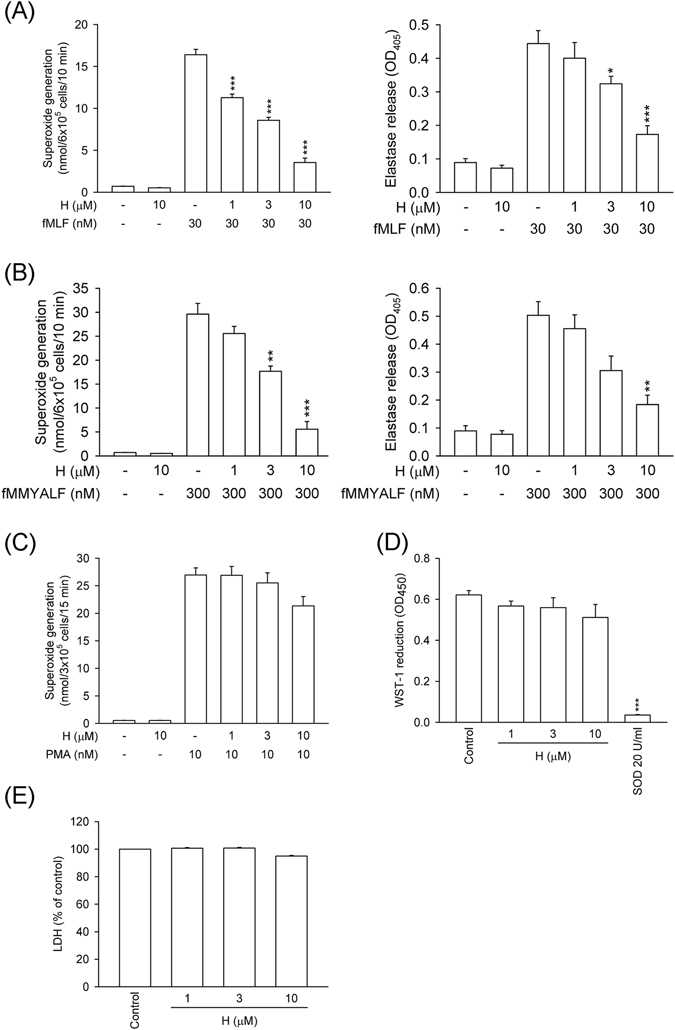
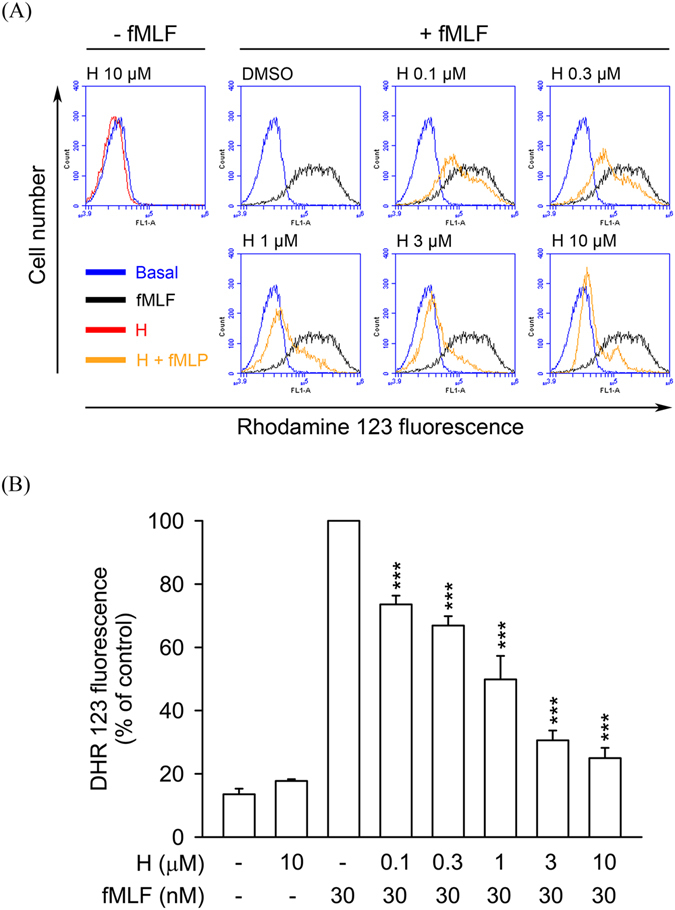
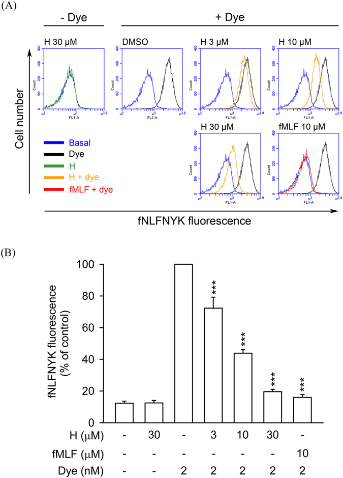
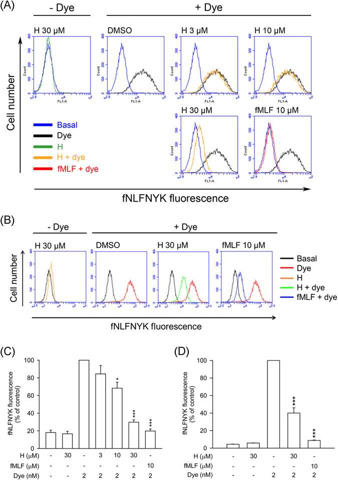
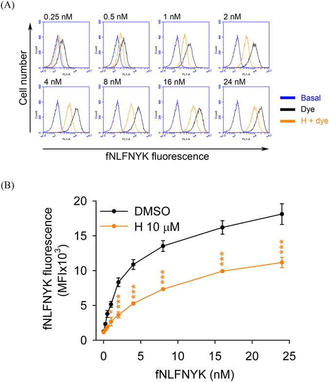
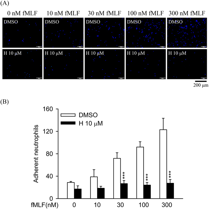
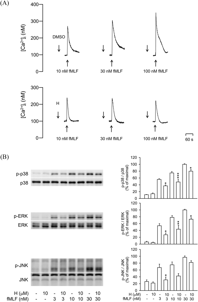
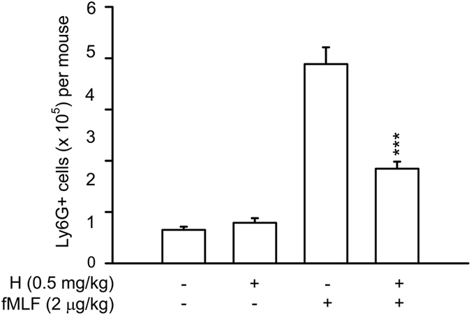
Formyl peptide receptor 1 (FPR1) mediates bacterial and mitochondrial N-formyl peptides-induced neutrophil activation. Therefore, FPR1 is an important therapeutic target for drugs to treat septic or sterile inflammatory diseases. Honokiol, a major bioactive compound of Magnoliaceae plants, possesses several anti-inflammatory activities. Here, we show that honokiol exhibits an inhibitory effect on FPR1 binding in human neutrophils. Honokiol inhibited superoxide anion generation, reactive oxygen species formation, and elastase release in bacterial or mitochondrial N-formyl peptides (FPR1 agonists)-activated human neutrophils. Adhesion of FPR1-induced human neutrophils to cerebral endothelial cells was also reduced by honokiol. The receptor-binding results revealed that honokiol repressed FPR1-specific ligand N-formyl-Nle-Leu-Phe-Nle-Tyr-Lys-fluorescein binding to FPR1 in human neutrophils, neutrophil-like THP-1 cells, and hFPR1-transfected HEK293 cells. However, honokiol did not inhibit FPR2-specific ligand binding to FPR2 in human neutrophils. Furthermore, honokiol inhibited FPR1 agonist-induced calcium mobilization as well as phosphorylation of p38 MAPK, ERK, and JNK in human neutrophils. In conclusion, our data demonstrate that honokiol may have therapeutic potential for treating FPR1-mediated inflammatory diseases.


Conflict of interest statement

The authors declare that they have no competing interests.

Figures

Figure 1
Figure 1
Honokiol inhibits superoxide anion generation and elastase release in FPR1-activated human neutrophils. Human neutrophils were incubated with 0.1% DMSO (as control) or honokiol (H; 1, 3, and 10 μM) for 5 min, and then activated by (A) fMLF (30 nM), (B) fMMYALF (300 nM), or (C) PMA (10 nM) for another 10 min. When fMLF and fMMYALF were used as activators, cells were primed by pre-incubation for 3 min with cytochalasin B (CB). Superoxide anion production was measured by ferricytochrome c reduction and elastase release was measured spectrophotometrically at 405 nm. (D) Xanthine oxidase was incubated with 0.1% DMSO (as control) or honokiol for 3 min, and then xanthine (0.1 mM) was added for 10 min. Reduction of WST-1 by cell-free xanthine/xanthine oxidase was measured spectrophotometrically at 450 nm. Superoxide dismutase (SOD; 20 U/ml) was used as a positive control. (E) Human neutrophils were incubated with 0.1% DMSO (as control) or honokiol for 15 min. Cytotoxicity was measured by assessing LDH release in cell-free medium. Total LDH release was determined by lysing cells with 0.1% Triton X-100 for 30 min at 37 °C. All data are expressed as the mean ± S.E.M. (n = 3–5). *P < 0.05, **P < 0.01, ***P < 0.001 compared with (A) fMLF, (B) fMMYALF, or (D) control (0.1% DMSO).
Figure 2
Figure 2
Honokiol inhibits ROS formation in fMLF-stimulated human neutrophils. Human neutrophils were labelled with DHR 123 (2 μM) and incubated with 0.1% DMSO (as control) or honokiol (H; 0.1–10 μM) for 5 min. Cells were activated using fMLF (30 nM) in the presence of CB (0.5 μg/ml) for a further 5 min. (A) Representative histograms showing typical fluorescent-activated cell sorting profiles for rhodamine 123 in unstimulated cells and in cells stimulated with fMLF in the absence or presence of honokiol at the concentration indicated. (B) Mean fluorescence intensity is shown as the mean ± S.E.M. (n = 5). ***P < 0.001 compared with fMLF alone.
Figure 3
Figure 3
Honokiol blocks fNLFNYK binding to FPR1 in human neutrophils. Human neutrophils were incubated with 0.1% DMSO (as control), honokiol (H; 3, 10, and 30 μM), or fMLF (10 μM) for 10 min and then labelled with FPR1-specific fluorescent ligand fNLFNYK (2 nM) for 20 min. (A) Representative histograms showing typical fluorescence in the absence or presence of fNLFNYK with honokiol or fMLF. (B) Mean fluorescence intensity is shown as the mean ± S.E.M. (n = 4). **P < 0.01; ***P < 0.001 compared with fluorescent dye alone.
Figure 4
Figure 4
Honokiol blocks fNLFNYK binding to FPR1 in FPR1-expressed neutrophil-like cells. (A) Dibutyryl cAMP-differentiated THP-1 or (B) hFPR1-transfected HEK-293 cells were incubated with 0.1% DMSO (as control), honokiol (H; 3, 10, or 30 μM), or fMLF (10 μM) for 10 min and then labelled with fNLFNYK (2 nM) for 20 min. (A,B) Representative histograms showing typical fluorescence in the absence or presence of fNLFNYK with honokiol or fMLF. (C and D) Mean fluorescence intensities of (A and B) are shown as the mean ± S.E.M. (n = 3). *P < 0.05; **P < 0.01; ***P < 0.001 compared with fluorescent dye alone.
Figure 5
Figure 5
Inhibitory effect of honokiol on dose–response curve of fNLFNYK in human neutrophils. Human neutrophils were incubated with 0.1% DMSO (as control) or honokiol (H; 10 μM) for 10 min before labelling with different concentrations of fNLFNYK (0.25–24 nM) for 20 min. (A) Representative histograms showing typical fluorescence of fNLFNYK in the absence or presence of honokiol. (B) Mean fluorescence intensity is shown as the mean ± S.E.M. (n = 5). *P < 0.05; **P < 0.01 compared with corresponding data.
Figure 6
Figure 6
Honokiol does not alter FPR1 mRNA expression and FPR2 binding effect in human neutrophils. (A) Human neutrophils were treated with 0.1% DMSO (as control), honokiol (10 and 30 μM), or fMLF (10 μM) at 4 °C for 30 min. Total RNAs were isolated with Trizol reagent and FPR1 mRNA levels were analyzed by quantitative PCR. (B) Human neutrophils were incubated with 0.1% DMSO (as control), honokiol (H; 10 μM), or WRW4 (1 μM) for 10 min and then labelled with FPR2-specific fluorescent ligand MMK-1F (200 nM) for 15 min. Representative histograms showing typical fluorescence in the absence or presence of MMK-1F with honokiol or WRW4. (C) Mean fluorescence intensity of (B) is shown. All data are expressed as the mean ± S.E.M. (n = 3 or 4). ***P < 0.001 compared with fluorescent dye alone.
Figure 7
Figure 7
Honokiol inhibits dose–response effects of fMLF in human neutrophils. Human neutrophils were incubated with 0.1% DMSO (as control) or honokiol (H; 10 μM) for 5 min before the addition of different concentrations of fMLF in the presence of CB. (A) Superoxide anion production was measured by assessing ferricytochrome c reduction. (B) Elastase release was measured spectrophotometrically at 405 nm. (C) Representative histograms of rhodamine 123 are shown. (D) Mean fluorescence intensities of (C) are shown. All data are expressed as the mean ± S.E.M. (n = 3–5). *P < 0.05; **P < 0.01; ***P < 0.001 compared with corresponding control.
Figure 8
Figure 8
Honokiol inhibits fMLF-induced adhesion of human neutrophils to endothelial cells (ECs). Hoechst-labelled neutrophils were incubated with 0.1% DMSO (as control) or honokiol (H; 10 μM) for 5 min before activation with different concentrations of fMLF and priming with CB. Activated neutrophils were then co-cultured with LPS-pre-activated ECs for 15 min. (A) Adherent neutrophils on ECs were detected using microscopy. Bar (bottom), 200 μm. (B) Adherent neutrophils were measured and are shown as the mean ± S.E.M. (n = 6). **P < 0.01 compared with control.
Figure 9
Figure 9
Honokiol inhibits Ca2+ mobilization and MAPKs phosphorylation in fMLF-activated human neutrophils. (A) Fluo-3/AM-labelled human neutrophils were incubated with 0.1% DMSO (as control) or honokiol (H; 10 μM) for 5 min. Cells were activated by fMLF and mobilization of Ca2+ was determined in real time by using a spectrofluorometer. Representative traces are shown (n = 7). (B) Human neutrophils were incubated with 0.1% DMSO (as the control) or honokiol (10 μM) for 5 min before stimulation with or without fMLF for another 25 s. Phosphorylation of p38, ERK, and JNK was analyzed by immunoblotting with antibodies against the phosphorylated and total form of each protein. All the Western blotting experiments were performed under the same condition. After transferred the blots onto nitrocellulose membranes, we immediately cropped the targeted blots according to referenced indicating markers, and then targeted proteins were immunoblotted with its specific monoclonal antibody. Data are normalized to the corresponding total protein level and expressed as mean ± S.E.M. relative to the mean maximal ratio (n = 4). *P < 0.05; **P < 0.01; ***P < 0.001 compared with corresponding control.
Figure 10
Figure 10
Honokiol inhibits neutrophils accumulation in the peritoneal cavity in fMLF-stimulated mice. Male BALB/c mice aged 7–8 weeks were pretreated by intraperitoneal injection of honokiol (0.5 mg/kg body weight) or vehicle alone (10% DMSO/normal saline) for 30 min, and then challenged with intraperitoneal injection of fMLF (2 μg/kg in normal saline) for 2 h. The peritoneal cells were harvested and stained with the Ly6G (Gr-1) rat anti-mouse, PE conjugated monoclonal antibody for 30 min at 4 °C. The number of Ly6G positive cells were detected by flow cytometry. Data are expressed as the mean ± S.E.M. (n = 7 in each group). ***P < 0.001 compared with fMLF.

Similar articles

Cited by

References

    1. Kolaczkowska E, Kubes P. Neutrophil recruitment and function in health and inflammation. Nat Rev Immunol. 2013;13:159–175. doi: 10.1038/nri3399. - DOI - PubMed
    1. von Andrian UH, et al. In vivo behavior of neutrophils from two patients with distinct inherited leukocyte adhesion deficiency syndromes. J Clin Invest. 1993;91:2893–2897. doi: 10.1172/JCI116535. - DOI - PMC - PubMed
    1. Angelillo-Scherrer A. Leukocyte-derived microparticles in vascular homeostasis. Circ Res. 2012;110:356–369. doi: 10.1161/CIRCRESAHA.110.233403. - DOI - PubMed
    1. Caielli S, Banchereau J, Pascual V. Neutrophils come of age in chronic inflammation. Curr Opin Immunol. 2012;24:671–677. doi: 10.1016/j.coi.2012.09.008. - DOI - PMC - PubMed
    1. Amulic B, Cazalet C, Hayes GL, Metzler KD, Zychlinsky A. Neutrophil function: from mechanisms to disease. Annu Rev Immunol. 2012;30:459–489. doi: 10.1146/annurev-immunol-020711-074942. - DOI - PubMed

Publication types

MeSH terms