. 2017 Jul 27;7(1):6665.
doi: 10.1038/s41598-017-06879-5.

Hydrogen sulphide donors selectively potentiate a green tea polyphenol EGCG-induced apoptosis of multiple myeloma cells

Affiliations

Hydrogen sulphide donors selectively potentiate a green tea polyphenol EGCG-induced apoptosis of multiple myeloma cells

Jaehoon Bae et al. Sci Rep. .

Abstract

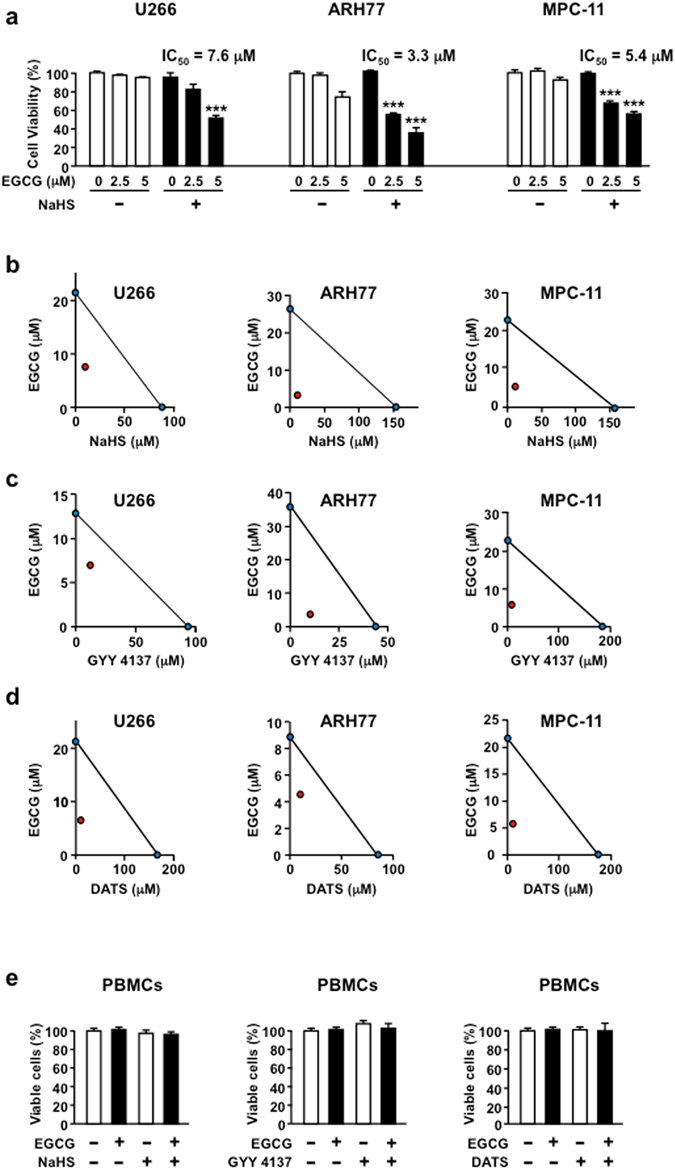
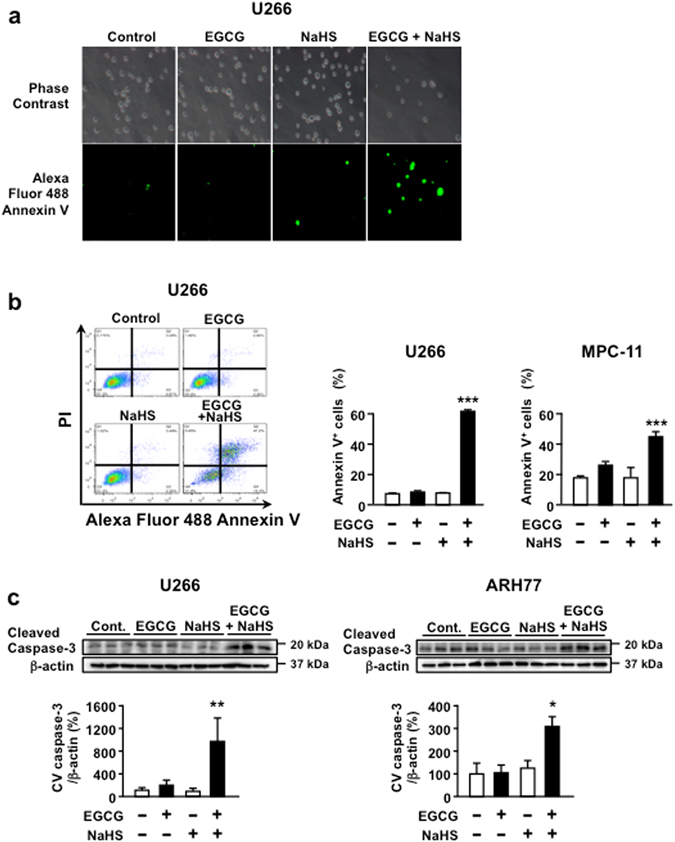
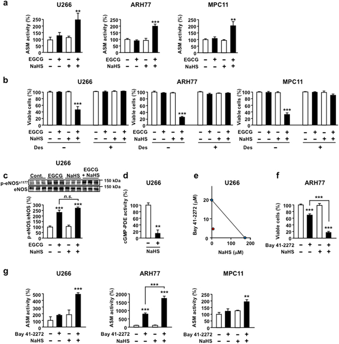
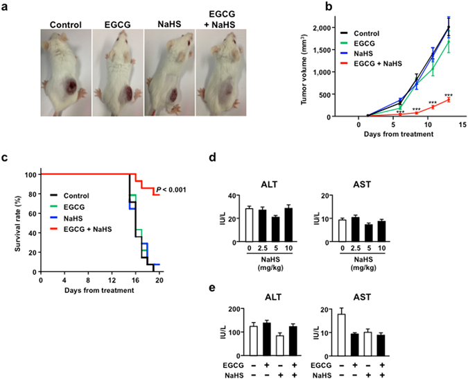
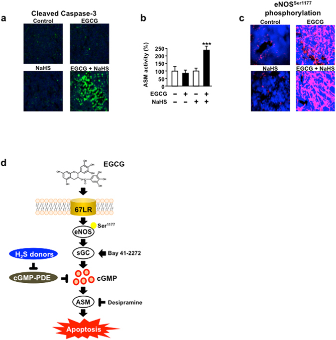
Hydrogen sulphide (H2S) is a colourless gas with the odour of rotten eggs and has recently been recognized as a signal mediator in physiological activities related with the regulation of homeostasis, the vascular system and the inflammatory system. Here we show that H2S donors, including sodium hydrogen sulphide (NaHS), GYY 4137 and diallyltrisulfide (DATS), synergistically enhanced the anti-cancer effect of a green tea polyphenol (-)-epigallocatechin-3-O-gallate (EGCG) against multiple myeloma cells without affecting normal cells. NaHS significantly potentiated the anti-cancer effect of EGCG and prolonged survival in a mouse xenograft model. In this mechanism, H2S enhanced apoptotic cell death through cyclic guanosine monophosphate (cGMP)/acid sphingomyelinase pathway induced by EGCG. Moreover, NaHS reduced the enzyme activity of cyclic nucleotide phosphodiesterase that is known as cGMP negative regulator. In conclusion, we identified H2S as a gasotransmitter that potentiates EGCG-induced cancer cell death.


Conflict of interest statement

The authors declare that they have no competing interests.

Figures

Figure 1
Figure 1
H2S donors potentiate EGCG cancer-specific cell death. (a) U266, ARH77 and MPC11 cells are cultured with or without NaHS (10 µM) and/or EGCG for 96 h, and viable cell numbers are measured. (bd) The combination effect of EGCG and several H2S donors in U266, ARH77 and MPC-11 cells are measured by isobologram analysis. (e) Normal PBMCs are treated with NaHS, GYY 4137 and DATS (10 µM) with or without EGCG (5 µM) for 96 h. Data are presented as mean ± SEM (n = 3). **P < 0.01, ***P < 0.001.
Figure 2
Figure 2
Combination of EGCG and NaHS induces apoptosis in MM cells. (a) U266 cells are treated or not treated with 5 μM EGCG in the presence or absence of 10 μM NaHS for 96 h and are observed under a florescence microscope. (b) Apoptotic cells are double stained with PI and Annexin V–Alexa Fluor 488 in U266 and MPC-11 cells. (c) U266 and ARH77 cells (human MM) are treated or not treated with 5 μM EGCG in the presence or absence of 10 μM NaHS for 72 h, and cleaved caspase-3 levels are assessed using western blot analysis. Cropped gels/blots are displayed. Full-length gels/blots are included in the Supplementary Information (Supplementary Fig. 7). Data are presented as mean ± SEM (n = 3). **P < 0.01, ***P < 0.001.
Figure 3
Figure 3
NaHS amplifies cGMP-dependent apoptosis signalling pathway. (a) The indicated cells are treated or not treated with 5 μM EGCG in the presence or absence of 10 μM NaHS and ASM activity is assessed using TLC analysis. (b) Multiple myeloma cell lines are pretreated or not pretreated with the ASM-specific inhibitor desipramine (Des; 5 μM) for 3 h and then are treated or not treated with EGCG (5 μM) and/or NaHS (10 µM) for 96 h. (c) U266 cells are treated or not treated with 5 μM EGCG in the presence or absence of 10 μM NaHS for 3 h, and phosphorylation of eNOS at Ser1177 is assessed using western blot analysis. Cropped gels/blots are displayed. Full-length gels/blots are included in the Supplementary Fig. 7. (d) Effect of 10 µM NaHS on cGMP-PDE enzyme activity in U266 cells. (e) Isobologram analysis of the effect of the combinations of Bay 41–2272 and NaHS. (f) ARH77 cells are treated or not treated with 5 μM Bay 41–2272 in the presence or absence of 10 μM NaHS for 96 h. (g) The indicated cells are treated or not treated with 5 μM Bay 41–2272 in the presence or absence of 10 μM NaHS, and ASM activity is assessed using TLC analysis. Data are presented as mean ± SEM (n = 3). **P < 0.01, ***P < 0.001.
Figure 4
Figure 4
NaHS potentiates the anti-MM effect of EGCG in vivo. (ac) MPC-11 cells are injected subcutaneously into female BALB/c mice, and mice (n = 14 per group) are administered i.p. injections of EGCG (15 mg/kg) and/or NaHS (10 mg/kg) every 2 days. Log-rank analyses of Kaplan–Meier curves are performed (n = 14). (d) Effect of the indicated NaHS concentration on the serum AST and ALT levels (n = 5). (e) Effect of the combination of EGCG and NaHS on the serum AST and ALT levels. Data are presented as mean ± SEM (n = 13). ***P < 0.001.
Figure 5
Figure 5
EGCG and NaHS amplify the tumour apoptosis in mouse xenograft model. (a) MPC-11 cells are injected subcutaneously into female BALB/c mice, and mice are administered i.p. injections of EGCG (15 mg/kg) and/or NaHS (10 mg/kg) every 2 days. Tumours are harvested and evaluated for cleaved caspase-3 (n = 6). (b) Tumours are harvested and evaluated for ASM activity (n = 13). (c) MPC11 cells are injected subcutaneously into female BALB/c mice, and mice are administered i.p. injections of EGCG (15 mg/kg) and/or NaHS (10 mg/kg) every 2 days. Tumours are harvested and evaluated for eNOS phosphorylation at Ser1177 (n = 6). (d) Schematic representation of the H2S donor potentiates EGCG-induced cell-death pathway. Data are presented as mean ± SEM. ***P < 0.001.

Similar articles

Cited by

References

    1. Naganuma T, et al. Green tea consumption and hematologic malignancies in Japan: the Ohsaki study. Am. J. Epidemiol. 2009;170:30–738. doi: 10.1093/aje/kwp187. - DOI - PubMed
    1. Tachibana H, Koga K, Fujimura Y, Yamada K. A receptor for green tea polyphenol EGCG. Nat. Struct. Mol. Biol. 2004;11:380–381. doi: 10.1038/nsmb743. - DOI - PubMed
    1. Tsukamoto S, et al. Oxygen partial pressure modulates 67-kDa laminin receptor expression, leading to altered activity of the green tea polyphenol, EGCG. FEBS Lett. 2012;586:3441–3447. doi: 10.1016/j.febslet.2012.07.065. - DOI - PubMed
    1. Kumazoe M, et al. 67-kDa laminin receptor increases cGMP to induce cancer-selective apoptosis. J. Clin. Invest. 2013;123:787–799. - PMC - PubMed
    1. Kumazoe M, et al. Vardenafil, a clinically available phosphodiesterase inhibitor, potentiates the killing effect of EGCG on CLL cells. Br. J. Haematol. 2015;168:610–613. doi: 10.1111/bjh.13135. - DOI - PubMed

Publication types

MeSH terms