. 2017 Jul 27;7(1):6723.
doi: 10.1038/s41598-017-06117-y.

Anti-AMPA GluA3 antibodies in Frontotemporal dementia: a new molecular target

Affiliations

Anti-AMPA GluA3 antibodies in Frontotemporal dementia: a new molecular target

B Borroni et al. Sci Rep. .

Erratum in

Abstract

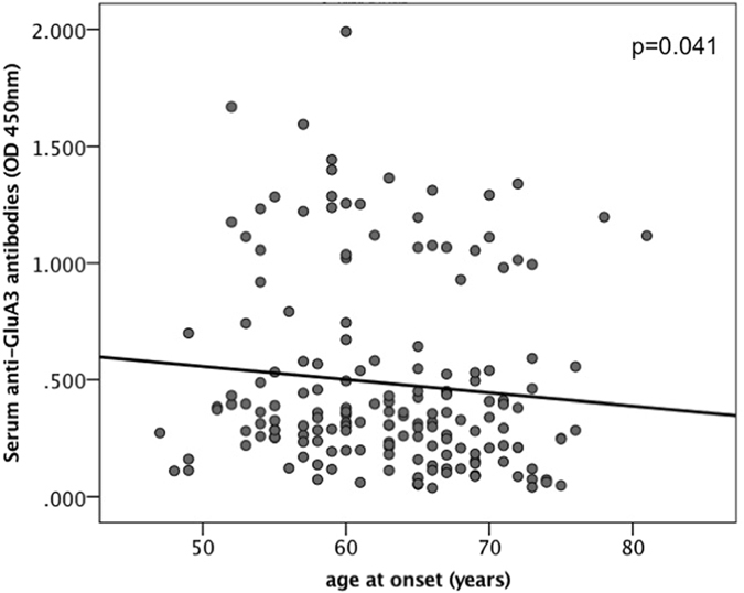
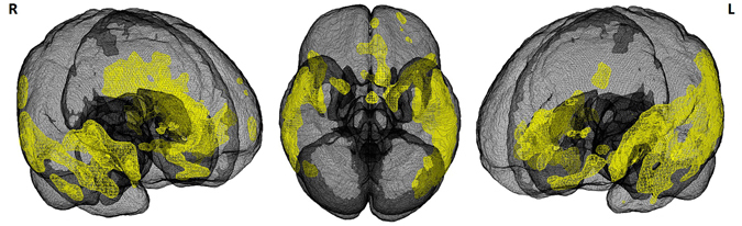
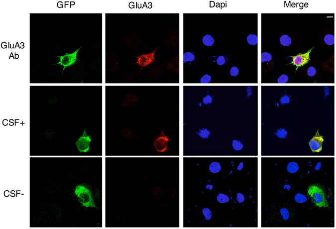
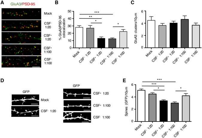
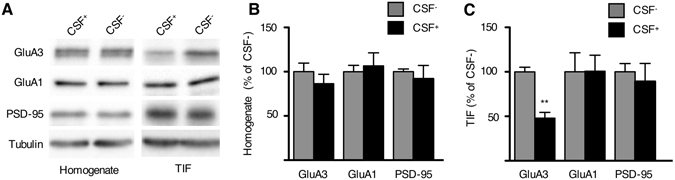
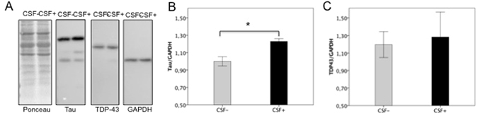
Frontotemporal Dementia (FTD) is a neurodegenerative disorder mainly characterised by Tau or TDP43 inclusions. A co-autoimmune aetiology has been hypothesised. In this study, we aimed at defining the pathogenetic role of anti-AMPA GluA3 antibodies in FTD. Serum and cerebrospinal fluid (CSF) anti-GluA3 antibody dosage was carried out and the effect of CSF with and without anti-GluA3 antibodies was tested in rat hippocampal neuronal primary cultures and in differentiated neurons from human induced pluripotent stem cells (hiPSCs). TDP43 and Tau expression in hiPSCs exposed to CSF was assayed. Forty-one out of 175 screened FTD sera were positive for the presence of anti-GluA3 antibodies (23.4%). FTD patients with anti-GluA3 antibodies more often presented presenile onset, behavioural variant FTD with bitemporal atrophy. Incubation of rat hippocampal neuronal primary cultures with CSF with anti-GluA3 antibodies led to a decrease of GluA3 subunit synaptic localization of the AMPA receptor (AMPAR) and loss of dendritic spines. These results were confirmed in differentiated neurons from hiPSCs, with a significant reduction of the GluA3 subunit in the postsynaptic fraction along with increased levels of neuronal Tau. In conclusion, autoimmune mechanism might represent a new potentially treatable target in FTD and might open new lights in the disease underpinnings.


Conflict of interest statement

The authors declare that they have no competing interests.

Figures

Figure 1
Figure 1
Inverse correlation between age at disease onset and anti-GluA3 antibody titer.
Figure 2
Figure 2
Brain atrophy in FTD patients with GluA3 antibodies. MRI scans were available in 18 FTD patients with anti-GluA3 antibodies (age: 62.4 ± 6.7, gender F: 38.9%) and were compared to 42 healthy controls (age: 61.0 ± 9.1, gender F: 66.7%) (SPM12 software package Wellcome Department of Imaging Neuroscience, London; http://www.fil.ion.ucl.ac.uk/spm). Statistical threshold was set at p < 0.05, Family Wise Error (FWE) corrected, with a minimum cluster size of 50 voxels.
Figure 3
Figure 3
Cell-based assay in heterologous COS7 cell. Confocal imaging of COS7 cells transfected with rat GluA3-GFP (green) and incubated with a GluA3 commercial antibody (upper panels), CSF+ (middle panels) or CSF− (lower panels) for the staining of GluA3 (red). Dapi was used to recognize cell nuclei (blue). Merge panels are shown on the right. Scale bar, 10 μm.
Figure 4
Figure 4
Neurobiological effect of GluA3 antibodies in primary rat neurons. (A) Fluorescence immunocytochemistry of GluA3 (green), PSD-95 (red) and MAP2 (blue) in DIV14 neurons treated with CSF with (CSF+) or without (CSF−) anti-GluA3 antibodies at two different dilutions (1:20 and 1:100) for 24 hours. Scale bar, 5 μm. (B) Bar graph representing the percentage of co-localization of GluA3 with PSD-95. Data are presented as mean ± s.e.m, n = 14, one-way ANOVA followed by Tukey as post hoc test; *p < 0.05, **p < 0.01, ***p < 0.001. (C) Bar graph representing the density of GluA3 positive cluster along dendrites. Data are presented as mean ± s.e.m., n = 14, one-way ANOVA followed by Tukey as post hoc test. (D) Confocal images of rat primary hippocampal neurons transfected at DIV8 with GFP and immunolabeled at DIV14 for GFP (green). Scale bar: 5 μm. (E), Bar graph showing the quantification of dendritic spine density. Data are presented as mean ± s.e.m., n = 6–8, one-way ANOVA followed by Tukey as post hoc test; *p < 0.05, **p < 0.01, ***p < 0.001.
Figure 5
Figure 5
Neurobiological effect of GluA3 antibodies in neurons differentiated from human iPSCs. (A) WB analysis from homogenates and triton-insoluble postsynaptic fractions (TIF) of neurons differentiated from human iPSC and incubated for 24 hours with CSF with (CSF+) or without (CSF−) anti-GluA3 antibodies (dilution 1:100). (B,C) The histogram shows the quantification of the expression levels of GluA3, GluA1 and PSD-95 in homogenate (B) and TIF (C), normalized on tubulin and expressed as % of CSF−. N = 5 ***p < 0.001, unpaired Student’s t-test.
Figure 6
Figure 6
Effect of GluA3 antibodies in neurons differentiated from human iPSc on Tau and TDP43 expression. (A) Representative WB analysis from homogenates of neurons differentiated from human iPSc and incubated for 24 hours with CSF with (CSF+) or without (CSF−) anti-GluA3 antibodies (dilution 1:100). (B,C) The histogram shows the quantification of the expression levels of Tau (B) and TDP43 (C), normalized on GAPDH; results are expressed as mean±standard errors (SEM), *p < 0.05, paired Student’s t-test.

References

    1. Gorno-Tempini ML, et al. Classification of primary progressive aphasia and its variants. Neurology. 2011;76:1006–1014. doi: 10.1212/WNL.0b013e31821103e6. - DOI - PMC - PubMed
    1. Rascovsky K, et al. Sensitivity of revised diagnostic criteria for the behavioural variant of frontotemporal dementia. Brain. 2011;134:2456–2477. doi: 10.1093/brain/awr179. - DOI - PMC - PubMed
    1. Rohrer JD, et al. Patterns of cortical thinning in the language variants of frontotemporal lobar degeneration. Neurology. 2009;72:1562–1569. doi: 10.1212/WNL.0b013e3181a4124e. - DOI - PMC - PubMed
    1. Bang J, Spina S, Miller BL. Frontotemporal dementia. Lancet. 2015;386:1672–1682. doi: 10.1016/S0140-6736(15)00461-4. - DOI - PMC - PubMed
    1. Borroni B, Padovani A. Dementia: a new algorithm for molecular diagnostics in FTLD. Nat. Rev. Neurol. 2013;9(5):241–242. doi: 10.1038/nrneurol.2013.72. - DOI - PubMed

Publication types

MeSH terms