

Activation of p47phox as a Mechanism of Bupivacaine-Induced Burst Production of Reactive Oxygen Species and Neural Toxicity
- PMID: 28751934
- PMCID: PMC5480047
- DOI: 10.1155/2017/8539026
Activation of p47phox as a Mechanism of Bupivacaine-Induced Burst Production of Reactive Oxygen Species and Neural Toxicity
Abstract
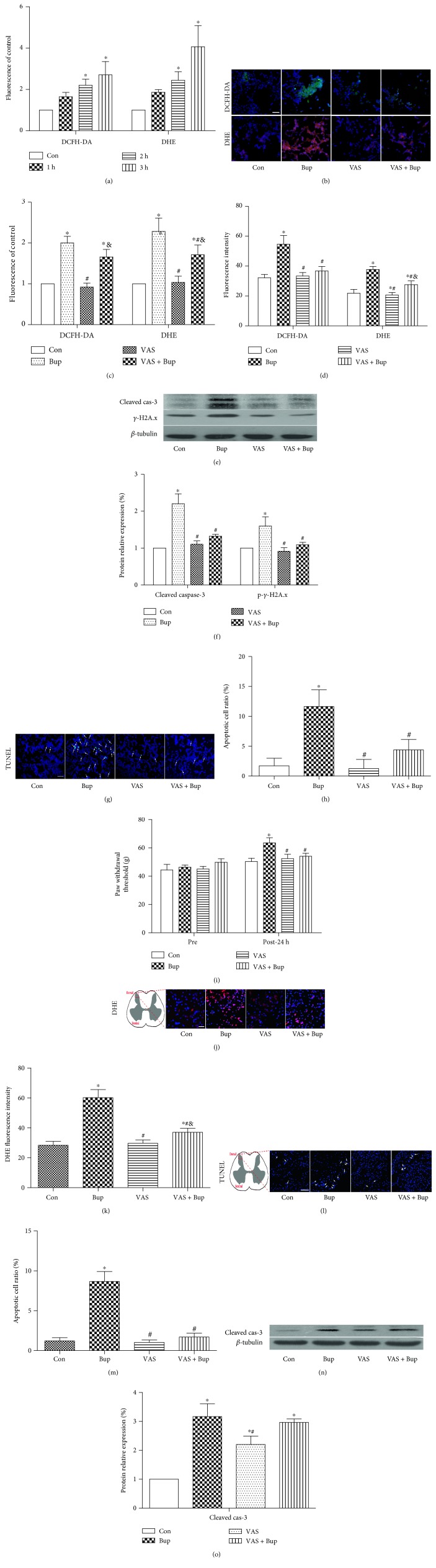
Bupivacaine has been shown to induce neurotoxicity through inducing excessive reactive oxygen species (ROS), but the underlying mechanism remains unclear. NOX2 is one of the most important sources of ROS in the nervous system, and its activation requires the membrane translocation of subunit p47phox. However, the role of p47phox in bupivacaine-induced neurotoxicity has not been explored. In our in vitro study, cultured human SH-SY5Y neuroblastoma cells were treated with 1.5 mM bupivacaine to induce neurotoxicity. Membrane translocation of p47phox was assessed by measuring the cytosol/membrane ratio of p47phox. The effects of the NOX inhibitor VAS2870 and p47phox-siRNA on bupivacaine-induced neurotoxicity were investigated. Furthermore, the effect of VAS2870 on bupivacaine-induced neurotoxicity was assessed in vivo in rats. All these changes were reversed by pretreatment with VAS2870 or transfection with p47phox-siRNA in SH-SY5Y cells. Similarly, pretreatment with VAS2870 attenuated bupivacaine-induced neuronal toxicity in rats. It is concluded that enhancing p47phox membrane translocation is a major mechanism whereby bupivacaine induced neurotoxicity and that pretreatment with VAS2870 or local p47phox gene knockdown attenuated bupivacaine-induced neuronal cell injury.
Figures


Similar articles
-
NADPH oxidase inhibitor improves outcome of mechanical reperfusion by suppressing hemorrhagic transformation.J Neurointerv Surg. 2017 May;9(5):492-498. doi: 10.1136/neurintsurg-2016-012377. Epub 2016 Apr 13. J Neurointerv Surg. 2017. PMID: 27075483
-
Ca(2+)-dependent p47phox translocation in hydroperoxide modulation of the alveolar macrophage respiratory burst.Am J Physiol. 1997 Nov;273(5):L1042-7. doi: 10.1152/ajplung.1997.273.5.L1042. Am J Physiol. 1997. PMID: 9374733
-
NADPH oxidase inhibitor VAS2870 prevents staurosporine-induced cell death in rat astrocytes.Radiol Oncol. 2019 Jan 19;53(1):69-76. doi: 10.2478/raon-2019-0002. Radiol Oncol. 2019. PMID: 30661061 Free PMC article.
-
Mechanistic study of mtROS-JNK-SOD2 signaling in bupivacaine-induced neuron oxidative stress.Aging (Albany NY). 2020 Jul 13;12(13):13463-13476. doi: 10.18632/aging.103447. Aging (Albany NY). 2020. PMID: 32658869 Free PMC article.
-
Organizers and activators: Cytosolic Nox proteins impacting on vascular function.Free Radic Biol Med. 2017 Aug;109:22-32. doi: 10.1016/j.freeradbiomed.2017.03.017. Epub 2017 Mar 21. Free Radic Biol Med. 2017. PMID: 28336130 Review.
Cited by
-
Implication of Nicotinamide Adenine Dinucleotide Phosphate (NADPH) Oxidase and Its Inhibitors in Alzheimer's Disease Murine Models.Antioxidants (Basel). 2021 Feb 2;10(2):218. doi: 10.3390/antiox10020218. Antioxidants (Basel). 2021. PMID: 33540840 Free PMC article. Review.
-
NOX2-Dependent Reactive Oxygen Species Regulate Formyl-Peptide Receptor 1-Mediated TrkA Transactivation in SH-SY5Y Cells.Oxid Med Cell Longev. 2019 Dec 2;2019:2051235. doi: 10.1155/2019/2051235. eCollection 2019. Oxid Med Cell Longev. 2019. PMID: 31871542 Free PMC article.
-
The roles of PARP-1 and XPD and their potential interplay in repairing bupivacaine-induced neuron oxidative DNA damage.Aging (Albany NY). 2021 Jan 20;13(3):4274-4290. doi: 10.18632/aging.202390. Epub 2021 Jan 20. Aging (Albany NY). 2021. PMID: 33495403 Free PMC article.
-
Monosialoganglioside protects against bupivacaine-induced neurotoxicity caused by endoplasmic reticulum stress in rats.Drug Des Devel Ther. 2019 Feb 19;13:707-718. doi: 10.2147/DDDT.S192225. eCollection 2019. Drug Des Devel Ther. 2019. PMID: 30858700 Free PMC article.
-
The role of NOX inhibitors in neurodegenerative diseases.IBRO Rep. 2019 Aug 1;7:59-69. doi: 10.1016/j.ibror.2019.07.1721. eCollection 2019 Dec. IBRO Rep. 2019. PMID: 31463415 Free PMC article. Review.
References
-
- Brull R., McCartney C. J., Chan V. W., El-Beheiry H. Neurological complications after regional anesthesia: contemporary estimates of risk. Anesthesia and Analgesia. 2007;104(4):965–974. doi: 10.1213/01.ane.0000258740.17193.ec. - DOI - PubMed
MeSH terms
Substances
LinkOut - more resources
Full Text Sources
Other Literature Sources
Miscellaneous
