. 2017:2017:9303158.
doi: 10.1155/2017/9303158. Epub 2017 Jun 7.

Chk1 Promotes DNA Damage Response Bypass following Oxidative Stress in a Model of Hydrogen Peroxide-Associated Ulcerative Colitis through JNK Inactivation and Chromatin Binding

Affiliations

Chk1 Promotes DNA Damage Response Bypass following Oxidative Stress in a Model of Hydrogen Peroxide-Associated Ulcerative Colitis through JNK Inactivation and Chromatin Binding

Kathrin Reissig et al. Oxid Med Cell Longev. 2017.

Abstract

Dysregulation of c-Jun N-terminal kinase (JNK) activation promoted DNA damage response bypass and tumorigenesis in our model of hydrogen peroxide-associated ulcerative colitis (UC) and in patients with quiescent UC (QUC), UC-related dysplasia, and UC-related carcinoma (UC-CRC), thereby adapting to oxidative stress. In the UC model, we have observed features of oncogenic transformation: increased proliferation, undetected DNA damage, and apoptosis resistance. Here, we show that Chk1 was downregulated but activated in the acute and quiescent chronic phases. In both phases, Chk1 was linked to DNA damage response bypass by suppressing JNK activation following oxidative stress, promoting cell cycle progression despite DNA damage. Simultaneously, activated Chk1 was bound to chromatin. This triggered histone acetylation and the binding of histone acetyltransferases and transcription factors to chromatin. Thus, chromatin-immobilized activated Chk1 executed a dual function by suppressing DNA damage response and simultaneously inducing chromatin modulation. This caused undetected DNA damage and increased cellular proliferation through failure to transmit the appropriate DNA damage signal. Findings in vitro were corroborated by chromatin accumulation of activated Chk1, Ac-H3, Ac-H4, and c-Jun in active UC (AUC) in vivo. Targeting chromatin-bound Chk1, GCN5, PCAF, and p300/CBP could be a novel therapeutic strategy to prevent UC-related tumor progression.


Figures

Figure 1
Figure 1
Chk1 is downregulated and activated in acute and quiescent chronic phase of experimental UC. (a) Immunoblot analysis of H2O2-treated HCEC. Lysates were analyzed by immunoblotting with Chk1, p-Chk1, and β-actin antibodies. β-actin served as loading control, and fold expression relative to HCEC is given below the blots. (b) Quantitative measurement of 8-OHdG in HCEC 24, 48, and 72 h after H2O2 treatment and in C10 cells 48 h following Chk1 siRNA transfection. (c) Immunoblot analysis of C-cell cultures. Lysates from C1-C10 cells and HCEC were immunoblotted with Chk1, p-Chk1, γ-H2AX, and β-actin antibodies. β-actin served as loading control, and fold expression relative to HCEC cells is given below the blots. γ-H2AX immunoblotting for C1 to C3 cells is already published in Poehlmann et al. [4]. (d) Cell cycle distribution of HCEC following Chk1 siRNA and control siRNA transfection and H2O2 treatment. The bar graphs represent the x-fold increase versus control siRNA as the mean ± S.E. of three independent experiments: P < 0.5, ∗∗P < 0.01.
Figure 2
Figure 2
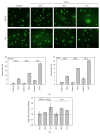
Comet assay analysis of H2O2-treated HCEC and C10 cells (a) and of C10 cells or H2O2-treated HCEC following Chk1 knockdown (b).
Figure 3
Figure 3
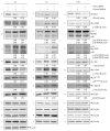
Chk1 promotes DNA damage response bypass by suppressing JNK activation. Immunoblot analysis of Chk1 siRNA-transfected and H2O2-treated HCEC 24, 48, and 72 h after H2O2 treatment. Lysates were analyzed by immunoblotting with Chk1, p-Chk1, JNK, p-JNK, c-Jun, p-c-Jun, p21WAF1, γ-H2AX, H2AX, and β-actin antibodies. β-actin served as loading control as marked (−, ∗, +, #, ◦, ~, and §), and fold expression relative to control siRNA is given below the blots.
Figure 4
Figure 4
Chk1 is a negative regulator of JNK activation in quiescent chronic phase of experimental UC. Immunoblot analysis of Chk1 siRNA-transfected C3, C5, and C10 cells 24 h after transfection. Lysates were analyzed by immunoblotting with Chk1, p-Chk1, JNK, p-JNK, c-Jun, p-c-Jun, p21WAF1, γ-H2AX, H2AX, and β-actin antibodies. β-actin served as loading control as marked (∗, +, #, ◦, and ~), and fold expression relative to control siRNA is given below the blots.
Figure 5
Figure 5
Chk1 downregulation contributes to defective maintenance of G2/M and mitotic spindle checkpoints. (a) Immunoblot analysis of Chk1 siRNA-transfected and H2O2-treated C10 cells 24 h after H2O2 treatment. Lysates were analyzed by immunoblotting with Chk1, p-Chk1, JNK, p-JNK, c-Jun, p-c-Jun, p21WAF1, γ-H2AX, H2AX, and β-actin antibodies. β-actin served as loading control as marked (∗, +, #, ◦, and ~), and fold expression relative to control siRNA is given below the blots. (b) Cell cycle distribution of C10 cells following Chk1 siRNA and control siRNA transfection and H2O2 treatment. Dashed lines contribute to cell cycle analysis with control siRNA transfection to mark increased G1 cell population and reduced G2/M arrest following Chk1 siRNA transfection. (c) Cell cycle distribution of HCEC, C5, and tetraploid C10 cells that were cultured under acidic conditions (10% CO2).
Figure 6
Figure 6
Increased expression of acetylated proteins and histone acetyltransferases in quiescent chronic phase. Immunoblot analysis of C1-C10 cells. Lysates were analyzed by immunoblotting with Ac-lysine and β-actin antibodies (a) or with Ac-H3, Ac-H4, p-H3T11, GCN5, PCAF, Ac-H3K9, and β-actin antibodies (b). β-actin served as loading control as marked (∗, +, #, ~, and −), and fold expression relative to HCEC is given below the blots.
Figure 7
Figure 7
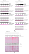
Acute and chronic DNA damage modulates Chk1 chromatin binding in vitro. (a) HCEC, H2O2-treated HCEC, C10 cells, and H2O2-treated C10 cells were fractionated, and chromatin-bound extracts were analyzed by immunoblotting using Chk1, p-Chk1, p-H3T11, GCN5, Ac-H3K9, Ac-H3, Ac-H4, PCAF, p300/CBP, ATF2, and c-Jun antibodies. Ponceau S staining served as loading control, and fold chromatin binding relative to HCEC is given below the blots. (b) Soluble nuclear and cytoplasmic extracts were analyzed by immunoblotting using Chk1 and p-Chk1 antibodies. Ponceau S staining served as loading control, and fold chromatin binding relative to HCEC is given below the blots.
Figure 8
Figure 8
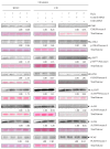
Acute, quiescent chronic, and active chronic oxidative stress induce chromatin modulation. HCEC and C10 cells were transfected with Chk1 siRNA or control siRNA, treated with H2O2, and subcellular fractionated. Chromatin-bound extracts were analyzed by immunoblotting using Chk1, p-Chk1, p-H3T11, GCN5, Ac-H3K9, Ac-H3, Ac-H4, and PCAF antibodies. Ponceau S staining served as loading control, and fold chromatin binding relative to control siRNA is given below the blots.
Figure 9
Figure 9
Activated Chk1, Ac-H3, Ac-H4, and c-Jun are chromatin-bound in AUC in vivo. Normal healthy colorectal mucosa (H1, H2, and H3) and inflamed tissue (I1–I4) was fractionated, and the chromatin-bound fractions were analyzed with p-Chk1, Ac-H3, and Ac-H4 antibodies (a) and with c-Jun, Ac-H3, and Ac-H4 antibodies (b). Ponceau S staining served as loading control. Localisations (a): healthy colorectal mucosa: H1, ascending colon; H2, descending colon; inflamed mucosa: I1, ascending colon; I2, ascending colon; I3, transverse colon; I4, descending colon. Localisations (b): healthy colorectal tissue: H1, ileum; H2; cecum; H3, ascending colon; inflamed mucosa: I1, transverse colon; I2, descending colon; I3, sigmoid colon; I4, rectum.
Figure 10
Figure 10
Proposed model for dual function of Chk1 in DNA damage response bypass in experimental UC. (a) On the one hand, Chk1 negatively regulates JNK activation, resulting in checkpoint override and reduced DNA damage response. On the other hand, activated Chk1 remains chromatin-bound and triggers acetylation and binding of transcription factors onto chromatin, leading to induction of proliferative genes and DNA damage response bypass. (b) In the acute phase, Chk1 and ATF2 dissociate from chromatin, while activated Chk1 accumulates on chromatin and phosphorylates H3 at T11. As a result, GCN5 is recruited and acetylates H3 at K9. Moreover, PCAF, Ac-H3, and c-Jun showed elevated chromatin binding. Consequently, cells underwent restrained cell cycle arrest. In the quiescent chronic phase, Chk1 and ATF2 again associate with chromatin despite DNA damage, further recruiting activated Chk1, Ac-H3, GCN5, PCAF, and Ac-H3K9, while p300/CBP is firstly recruited to chromatin. These chromatin changes resulted in increased proliferation and undetected DNA damage. In active chronic phase, chromatin binding of Chk1 and activated Chk1 is maintained, while levels of GCN5 Ac-H3K9, Ac-H3, Ac-H4, PCAF, p300/CBP, ATF2, and c-Jun are increased. As a result, cells underwent reversible cell cycle arrest and apoptosis resistance. Arrows indicate the appropriate relationship.

Similar articles

Cited by

References

    1. Arai N., Mitomi H., Ohtani Y., Igarashi M., Kakita A., Okayasu I. Enhanced epithelial cell turnover associated with p53 accumulation and high p21WAF1/CIP1 expression in ulcerative colitis. Modern Pathology. 1999;12(6):604–611. - PubMed
    1. Araki Y., Sugihara H., Hattori T. In vitro effects of dextran sulfate sodium on a Caco-2 cell line and plausible mechanisms for dextran sulfate sodium-induced colitis. Oncology Reports. 2006;16(6):1357–1362. doi: 10.3892/or.16.6.1357. - DOI - PubMed
    1. MacFie T. S., Poulsom R., Parker A., et al. DUOX2 and DUOXA2 form the predominant enzyme system capable of producing the reactive oxygen species H2O2 in active ulcerative colitis and are modulated by 5-aminosalicylic acid. Inflammatory Bowel Diseases. 2014;20(3):514–524. doi: 10.1097/01.MIB.0000442012.45038.0e. - DOI - PubMed
    1. Poehlmann A., Reissig K., Just A., et al. Non-apoptotic function of caspases in a cellular model of hydrogen peroxide-associated colitis. Journal of Cellular and Molecular Medicine. 2013;17(7):901–913. doi: 10.1111/jcmm.12079. - DOI - PMC - PubMed
    1. Poehlmann A., Reissig K., Schonfeld P., et al. Repeated H2O2 exposure drives cell cycle progression in an in vitro model of ulcerative colitis. Journal of Cellular and Molecular Medicine. 2013;17(12):1619–1631. doi: 10.1111/jcmm.12150. - DOI - PMC - PubMed

MeSH terms