

Characterization of Soft Contact Lens Fitting Using Ultra-Long Scan Depth Optical Coherence Tomography
- PMID: 28751981
- PMCID: PMC5471562
- DOI: 10.1155/2017/5763172
Characterization of Soft Contact Lens Fitting Using Ultra-Long Scan Depth Optical Coherence Tomography
Abstract
Objectives: To evaluate the centration and movement of soft contact lenses and to verify the repeatability of two repeated measurements of the lens centration and movement using ultra-long scan depth optical coherence tomography (UL-OCT).
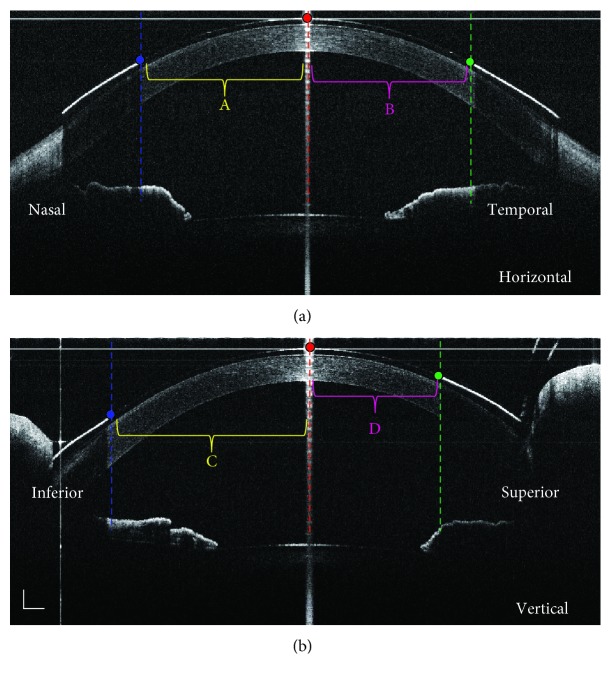
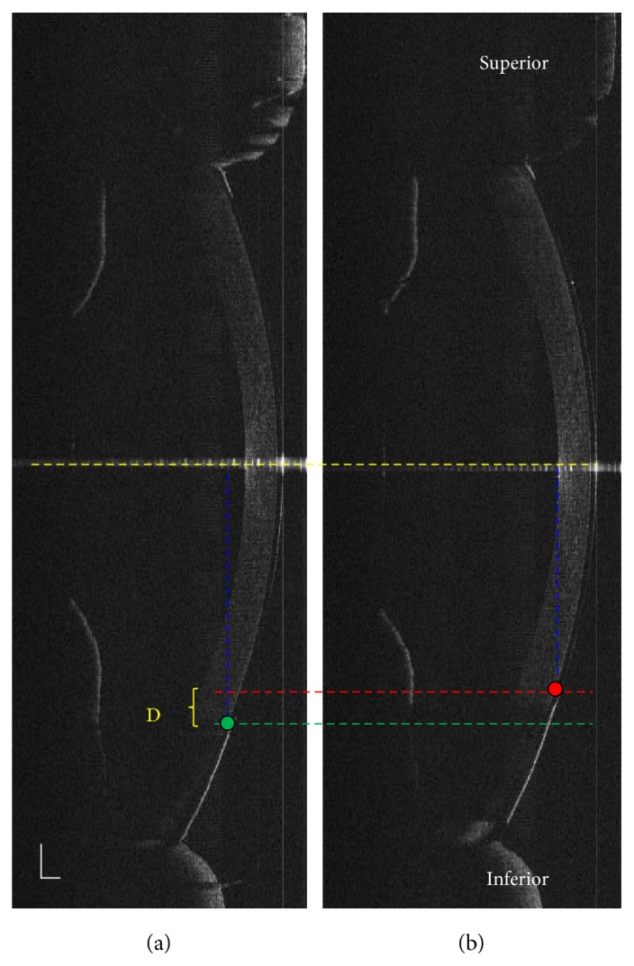
Methods: A 1-day Acuvue® Define™ lens was tested on both eyes of 10 subjects (5 males and 5 females; mean age, 31.6 years). The centration and blink-induced movement of the contact lens were measured using UL-OCT at 5 min and 30 min after insertion. The measurements were repeated once at each checkpoint.
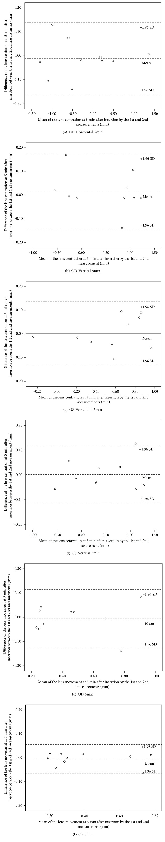
Results: Good repeatability was found in the lens centration and movement between the two repeated measurements at either checkpoint. The values of the lens movement were 0.457 ± 0.248 mm and 0.402 ± 0.229 mm at 5 min and decreased to 0.197 ± 0.065 mm and 0.211 ± 0.110 mm at 30 min after insertion for the right and left eyes, respectively (P < 0.05).
Conclusions: The custom-built UL-OCT presented good repeatability of centration and movement in Define lenses at 5 min and 30 min after insertion. Most of the lenses were centered temporal and inferior to the cornea during the first 30 min wearing period. Compared with 5 min after insertion, the lens was centered better and exhibited less movement at 30 min.
Figures


Similar articles
-
Characterization of soft contact lens edge fitting using ultra-high resolution and ultra-long scan depth optical coherence tomography.Invest Ophthalmol Vis Sci. 2011 Jun 9;52(7):4091-7. doi: 10.1167/iovs.10-6507. Invest Ophthalmol Vis Sci. 2011. PMID: 21372023 Free PMC article.
-
Fitting characteristics of 1-day and 14-day Acuvue disposable contact lenses.Optom Vis Sci. 1996 Dec;73(12):750-3. doi: 10.1097/00006324-199612000-00006. Optom Vis Sci. 1996. PMID: 9002092 Clinical Trial.
-
Objective analysis of contact lens fit.Cont Lens Anterior Eye. 2015 Jun;38(3):163-7. doi: 10.1016/j.clae.2015.01.006. Epub 2015 Feb 25. Cont Lens Anterior Eye. 2015. PMID: 25726510 Clinical Trial.
-
Simplified recording of soft contact lens fit.Cont Lens Anterior Eye. 2009 Feb;32(1):37-42. doi: 10.1016/j.clae.2008.12.004. Epub 2009 Jan 22. Cont Lens Anterior Eye. 2009. PMID: 19167261
-
Optical coherence tomography and scleral contact lenses: clinical and research applications.Clin Exp Optom. 2019 May;102(3):224-241. doi: 10.1111/cxo.12814. Epub 2018 Jul 30. Clin Exp Optom. 2019. PMID: 30062745 Review.
Cited by
-
Safety and Efficacy of a New Water Gradient Biomimetic Monthly Replacement Spherical Contact Lens Material (Lehfilcon A).Clin Ophthalmol. 2022 Aug 30;16:2873-2884. doi: 10.2147/OPTH.S362926. eCollection 2022. Clin Ophthalmol. 2022. PMID: 36065354 Free PMC article. Clinical Trial.
References
-
- Wichterle O., Lim D. Hydrophilic gels for biological use. Nature. 1962;185(4706):117–118. doi: 10.1038/185117a0. - DOI
-
- Spisato P. The soft lens story. Contact Lens Spectrum. 1987;2:65–77.
-
- Pritchard N., Fonn D., Weed K. Ocular and subjective responses to frequent replacement of daily wear soft contact lenses. The CLAO Journal. 1996;22(1):53–59. - PubMed
-
- Morgan P. Healthcheck on the contact lens market. The Optician. 2003;226(5908):32–33.
Grants and funding
LinkOut - more resources
Full Text Sources
Other Literature Sources
