

Ultrasound Imaging of Cystic Nephroma
- PMID: 28752022
- PMCID: PMC5519771
- DOI: 10.15586/jkcvhl.2017.79
Ultrasound Imaging of Cystic Nephroma
Abstract
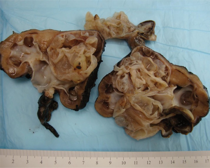
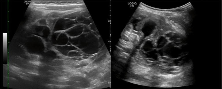
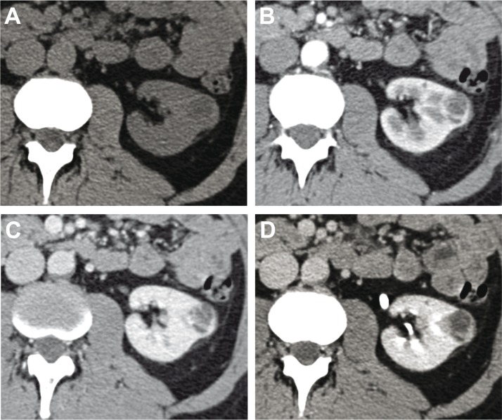
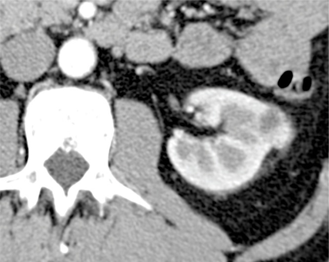
Cystic nephroma is a rare, benign multicystic lesion of the kidney. This tumor occurs both in children and in adults. In children, it is highly prevalent in males; in adults, it is more frequent in women. The term "cystic nephroma" represents two apparently different entities: pediatric cystic nephroma, a benign form thought to originate from metanephric tissue, and adult cystic nephroma, considered as a lesion of mixed epithelial stromal tumor. The clinical presentation may be a palpable mass or nonspecific symptoms such as abdominal pain, hematuria, and urinary tract infections. In this review, we summarize the ultrasound imaging features of cystic nephroma and describe the characteristics of the most common renal cystic lesions and the differential diagnosis of cystic nephroma with other renal cystic lesions.
Keywords: DICER1; cystic nephroma; cystic renal cell carcinoma; mixed epithelial stromal tumor; renal cystic lesions.
Figures
Similar articles
-
A MEST up classification? Review of the re-classification of mixed epithelial and stromal tumor and adult cystic nephroma for the abdominal radiologist.Abdom Radiol (NY). 2021 Feb;46(2):696-702. doi: 10.1007/s00261-020-02687-0. Epub 2020 Aug 5. Abdom Radiol (NY). 2021. PMID: 32757072 Review.
-
Adult multilocular cystic nephroma: Report of six cases with clinical, radio-pathologic correlation and review of literature.Urol Ann. 2013 Jan;5(1):13-7. doi: 10.4103/0974-7796.106958. Urol Ann. 2013. PMID: 23662002 Free PMC article.
-
Pediatric Cystic Nephroma Is Morphologically, Immunohistochemically, and Genetically Distinct From Adult Cystic Nephroma.Am J Surg Pathol. 2017 Apr;41(4):472-481. doi: 10.1097/PAS.0000000000000816. Am J Surg Pathol. 2017. PMID: 28177962 Free PMC article.
-
Mixed epithelial and stromal tumor of the kidney and cystic nephroma share overlapping features: reappraisal of 15 lesions.Arch Pathol Lab Med. 2006 Jan;130(1):80-5. doi: 10.5858/2006-130-80-MEASTO. Arch Pathol Lab Med. 2006. PMID: 16390243
-
[Cystic nephroma. Report of two cases and bibliographic review.].Arch Esp Urol. 2016 Dec;69(10):711-715. Arch Esp Urol. 2016. PMID: 28042792 Review. Spanish.
Cited by
-
A MEST up classification? Review of the re-classification of mixed epithelial and stromal tumor and adult cystic nephroma for the abdominal radiologist.Abdom Radiol (NY). 2021 Feb;46(2):696-702. doi: 10.1007/s00261-020-02687-0. Epub 2020 Aug 5. Abdom Radiol (NY). 2021. PMID: 32757072 Review.
-
Imaging of Kidney Cysts and Cystic Kidney Diseases in Children: An International Working Group Consensus Statement.Radiology. 2019 Mar;290(3):769-782. doi: 10.1148/radiol.2018181243. Epub 2019 Jan 1. Radiology. 2019. PMID: 30599104 Free PMC article.
-
Adult cystic nephroma masquerading urothelial carcinoma of a calyceal diverticulum.BMJ Case Rep. 2021 Oct 19;14(10):e245270. doi: 10.1136/bcr-2021-245270. BMJ Case Rep. 2021. PMID: 34667043 Free PMC article.
-
Case Report: A rare case of mixed epithelial and stromal tumor of the kidney in an adolescent: imaging findings and literature review.Front Pediatr. 2025 May 8;13:1550425. doi: 10.3389/fped.2025.1550425. eCollection 2025. Front Pediatr. 2025. PMID: 40406359 Free PMC article.
-
Case - Mixed epithelial and stromal tumours: A rare pediatric renal tumour.Can Urol Assoc J. 2019 Jan;13(1):E22-E24. doi: 10.5489/cuaj.5425. Can Urol Assoc J. 2019. PMID: 30059281 Free PMC article. No abstract available.
References
-
- Bonsib SM. Cystic nephroma. Mixed epithelial and stromal tumor. In: Eble JN, Sauter G, Epstein JL, Sesterhenn IA, editors. Pathology and genetics of tumors of the urinary system and male genital organs; WHO classification of tumours. Lyon: IARC Press; 2004. p. 76.
-
- Mohanty D, Jain BK, Agrawal V, Gupta A. Cystic nephroma: A diagnostic dilemma. Saudi J Kidney Dis Transpl. 2010. May;21(3):518–20. - PubMed
-
- Eble JN. Cystic nephroma and cystic partially differentiated nephroblastoma: Two entities or one? Adv Anat Pathol. 1994. Sep-Oct;1(2):99–102. http://dx.doi.org/10.1097/00125480-199409000-00007 - DOI
-
- Van Den Hoek J, De Krijger R, Van De Ven K, Lequin M, Van Den Heuvel-Eibrink MM. Cystic nephroma, cystic partially differentiated nephroblastoma and cystic Wilms’ tumor in children: A spectrum with therapeutic dilemmas. Urol Int. 2009. January;82(1):65–70. http://dx.doi.org/10.1159/000176028 - DOI - PubMed
-
- Joshi VV, Beckwith JB. Multilocular cyst of the kidney (cystic nephroma) and cystic, partially differentiated nephroblastoma. Terminology and criteria for diagnosis. Cancer. 1989. July;64(2):466–79. http://dx.doi.org/10.1002/1097-0142(19890715)64:2%3C466::AID-CNCR2820640... - DOI - PubMed
Publication types
LinkOut - more resources
Full Text Sources
Other Literature Sources