Clinical Trial
. 2017:2017:2321916.
doi: 10.1155/2017/2321916. Epub 2017 May 28.

Automated Image Analysis of HER2 Fluorescence In Situ Hybridization to Refine Definitions of Genetic Heterogeneity in Breast Cancer Tissue

Affiliations
Clinical Trial

Automated Image Analysis of HER2 Fluorescence In Situ Hybridization to Refine Definitions of Genetic Heterogeneity in Breast Cancer Tissue

Gedmante Radziuviene et al. Biomed Res Int. 2017.

Abstract

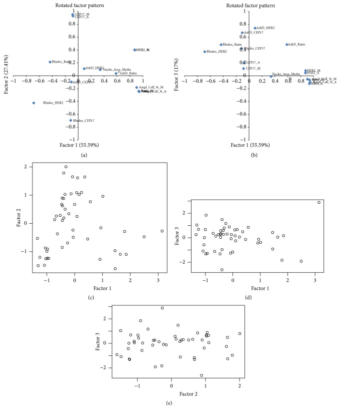
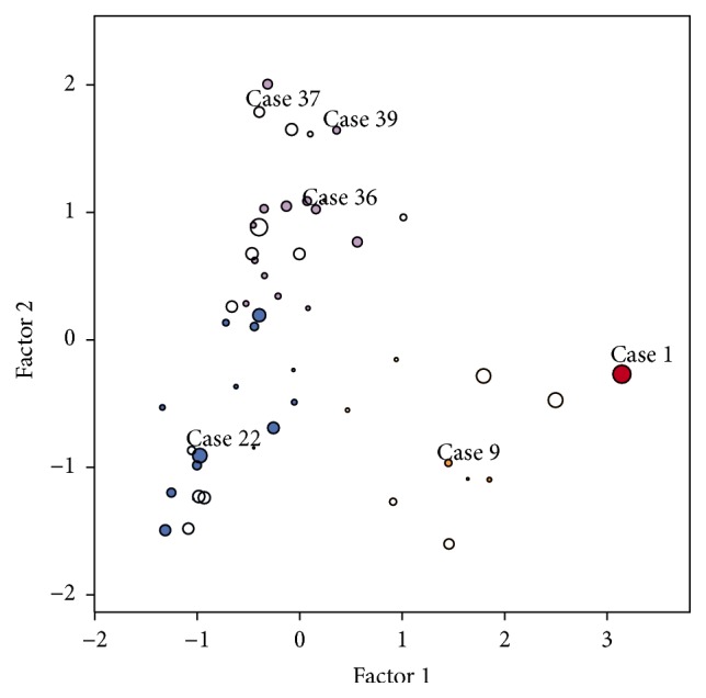
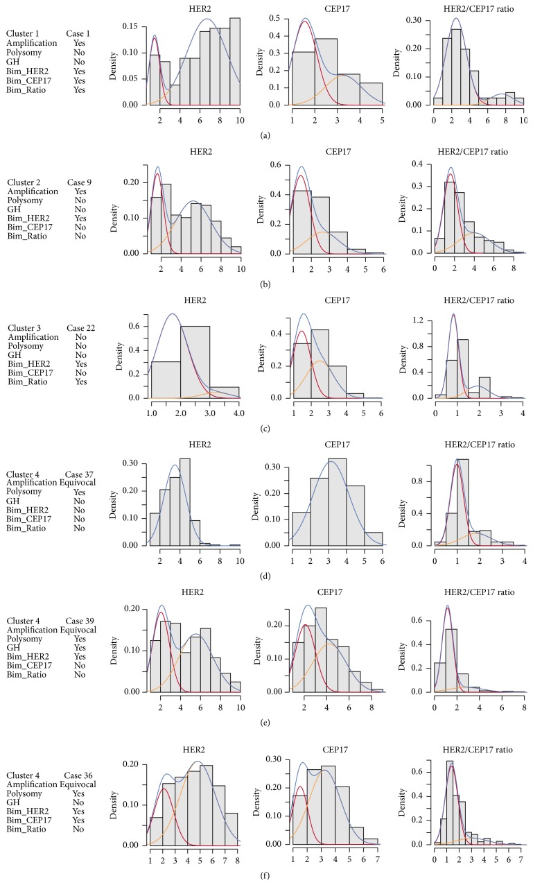
Human epidermal growth factor receptor 2 gene- (HER2-) targeted therapy for breast cancer relies primarily on HER2 overexpression established by immunohistochemistry (IHC) with borderline cases being further tested for amplification by fluorescence in situ hybridization (FISH). Manual interpretation of HER2 FISH is based on a limited number of cells and rather complex definitions of equivocal, polysomic, and genetically heterogeneous (GH) cases. Image analysis (IA) can extract high-capacity data and potentially improve HER2 testing in borderline cases. We investigated statistically derived indicators of HER2 heterogeneity in HER2 FISH data obtained by automated IA of 50 IHC borderline (2+) cases of invasive ductal breast carcinoma. Overall, IA significantly underestimated the conventional HER2, CEP17 counts, and HER2/CEP17 ratio; however, it collected more amplified cells in some cases below the lower limit of GH definition by manual procedure. Indicators for amplification, polysomy, and bimodality were extracted by factor analysis and allowed clustering of the tumors into amplified, nonamplified, and equivocal/polysomy categories. The bimodality indicator provided independent cell diversity characteristics for all clusters. Tumors classified as bimodal only partially coincided with the conventional GH heterogeneity category. We conclude that automated high-capacity nonselective tumor cell assay can generate evidence-based HER2 intratumor heterogeneity indicators to refine GH definitions.


Figures

Figure 1
Figure 1
Distribution of tumors with regard to genetic heterogeneity and amplification guidelines. (a) Percentage of amplified cells plotted for MP and AD values, Amp_Cell_%_M and Amp_Cell_%_A, respectively. Dashed line marks identity line. AD overestimates low MP values (crosses) whereas it underestimates MP values in the range >28 (circles); (b) HER2_M plotted against Ratio_M with cut-offs for amplification by ASCO/CAP 2013 guidelines shown by grey lines. Cases marked with crosses are overestimated cases from (a); (c) Cell_Amp_%_M plotted against Ratio_M, horizontal lines at 5% and 50% mark cut-off values for determining GH cases. Note the lack of cases in the 3–28% range; (d) HER2_A plotted against RATIO_A, amplification cut-off marked in grey; (e) Amp_Cell_%_A plotted against RATIO_A. Summary: MP: 13 positive, 21 equivocal, 16 negative, and 8 GH cases; AD: 7 positive, 3 equivocal, 40 negative, and 36 GH cases.
Figure 2
Figure 2
Rotated factor pattern of the indicators obtained by the manual and automated HER2 FISH procedure; n = 50. The loadings of (a) factors 1 and 2, (b) factors 1 and 3, and the factor scores (c, d, e) are plotted. Factor 1, amplification; factor 2, polysomy; factor 3, bimodality. Cell_Amp_%_M: percentage of amplified cells detected by manual procedure, calculated from HER2/CEP17 ratio. Cell_Amp_%_A: percentage of amplified cells detected by automated procedure, calculated from HER2/CEP17 ratio. HER2_A, HER2_M - HER2 copy number detected by automated and manual procedures, respectively. CEP17_A, CEP17_M - CEP17 copy number detected by automated and manual procedures, respectively. Ratio_A, Ratio_M - HER2/CEP17 ratio detected by automated and manual procedures, respectively. AshD_Ratio, AshD_HER2, and AshD_CEP17: Ashman's D indicator calculated for HER2/CEP17, HER2, and CEP17 automated data. BIndex_HER2, BIndex_CEP17, and BIndex_Ratio: bimodality indices calculated for HER2, CEP17, and HER2/CEP17 automated data.
Figure 3
Figure 3
A bubble plot of the clusters obtained from the factor 1, 2, and 3 scores. Cluster colors: Cluster 1, red; Cluster 2, orange; Cluster 3, blue; and Cluster 4, purple. Bubble size represents factor 3 (bimodality); center is empty for negative and filled for positive values. Numbers indicate cluster examples depicted in Figure 4.
Figure 4
Figure 4
Examples of the tumor cases from the clusters extracted from the automated HER2 FISH data. Histograms of HER2, CEP17, and HER2/CEP17 with Gaussian curves are presented from the cases labeled in the Figure 3. Amplification, polysomy, and genetic heterogeneity categories are based on the conventional manual procedure results. Bim_HER2, Bim_CEP17, and Bim_Ratio represent bimodality categories based on Ashman's D > 2 criterion.

References

    1. Slamon D. J., Clark G. M., Wong S. G., Levin W. J., Ullrich A., McGuire W. L. Human breast cancer: correlation of relapse and survival with amplification of the HER-2/neu oncogene. Science. 1987;235(4785):177–182. - PubMed
    1. Gutierrez C., Schiff R. HER2: biology, detection, and clinical implications. Archives of Pathology & Laboratory Medicine. 2011;135(1):55–62. - PMC - PubMed
    1. Wolff A. C., Hammond M. E., Hicks D. G. Recommendations for human epidermal growth factor receptor 2 testing in breast cancer: American Society of Clinical Oncology/College of American Pathologists clinical practice guideline update. Journal Clinical Oncology. 2013;31(31):3997–4013. doi: 10.1200/JCO.2013.50.9984. - DOI - PubMed
    1. Hicks D. G., Kulkarni S. Trastuzumab as adjuvant therapy for early breast cancer: the importance of accurate human epidermal growth factor receptor 2 testing. Archives of Pathology & Laboratory Medicine. 2008;132(6):1008–1015. - PubMed
    1. Krell J., James C. R., Shah D., et al. Human epidermal growth factor receptor 2-positive breast cancer relapsing post-adjuvant trastuzumab: pattern of recurrence, treatment and outcome. Clinical Breast Cancer. 2011;11(3):153–160. doi: 10.1016/j.clbc.2011.03.012. - DOI - PubMed

Publication types

MeSH terms