. 2017 Dec;190(3):304-314.
doi: 10.1111/cei.13018. Epub 2017 Sep 7.

Intranasal delivery of dexamethasone efficiently controls LPS-induced murine neuroinflammation

Affiliations

Intranasal delivery of dexamethasone efficiently controls LPS-induced murine neuroinflammation

G Meneses et al. Clin Exp Immunol. 2017 Dec.

Abstract

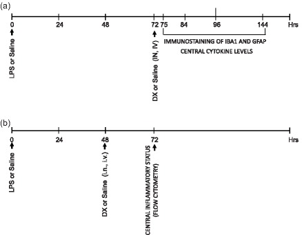
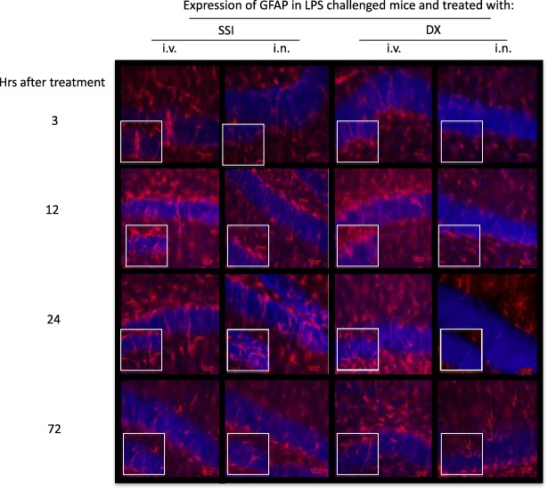
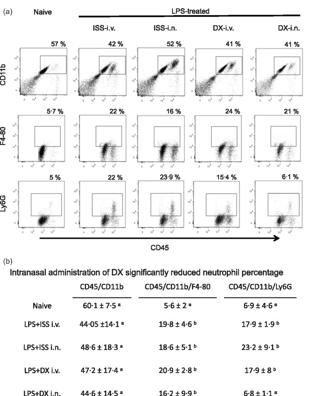
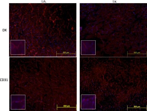
Neuroinflammation is the hallmark of several infectious and neurodegenerative diseases. Synthetic glucocorticoids (GCs) are the first-line immunosuppressive drugs used for controlling neuroinflammation. A delayed diffusion of GCs molecules and the high systemic doses required for brain-specific targeting lead to severe undesirable effects, particularly when lifelong treatment is required. Therefore, there is an urgent need for improving this current therapeutic approach. The intranasal (i.n.) route is being employed increasingly for drug delivery to the brain via the olfactory system. In this study, the i.n. route is compared to the intravenous (i.v.) administration of GCs with respect to their effectiveness in controlling neuroinflammation induced experimentally by systemic lipopolysaccharide (LPS) injection. A statistically significant reduction in interleukin (IL)-6 levels in the central nervous system (CNS) in the percentage of CD45+ /CD11b+ /lymphocyte antigen 6 complex locus G6D [Ly6G+ and in glial fibrillary acidic protein (GFAP) immunostaining was observed in mice from the i.n.-dexamethasone (DX] group compared to control and i.v.-DX-treated animals. DX treatment did not modify the percentage of microglia and perivascular macrophages as determined by ionized calcium binding adaptor molecule 1 (Iba1) immunostaining of the cortex and hippocampus. The increased accumulation of DX in brain microvasculature in DX-i.n.-treated mice compared with controls and DX-IV-treated animals may underlie the higher effectiveness in controlling neuroinflammation. Altogether, these results indicate that IN-DX administration may offer a more efficient alternative than systemic administration to control neuroinflammation in different neuropathologies.

Keywords: LPS; glucocorticoids; inflammation; intranasal route; neuroinflammation.


Figures

Figure 1
Figure 1
Experimental design. (a) Groups of five to six mice received lipopolysaccharide (LPS) or saline injected intraperitoneally (i.p.) (day 0). Three days later, mice were treated with saline or dexamethasone (DX), delivered intranasally (i.n.) or intravenously (i.v.). The expression of ionized calcium binding adaptor molecule 1 (Iba1) and glial fibrillary acidic protein (GFAP) was studied 3, 12, 24 and 72 h later by immunostaining. Central cytokine levels were evaluated by enzyme‐linked immunosorbemt assay (ELISA) 24 h after DX treatment. (b) The percentage of central cell populations was measured 24 h after DX treatment applied 2 days after LPS injection.
Figure 2
Figure 2
Overview of subregions, including cornu ammonis (CA1 and CA2). Two regions of the cortex were evaluated (R1 and R2). [Colour figure can be viewed at wileyonlinelibrary.com]
Figure 3
Figure 3
Effect of dexamethasone (DX) on glial fibrillary acidic protein (GFAP) expression in lipopolysaccharide (LPS)‐treated mice. Representative immunofluorescence of 30 μm coronal sections of mouse brain of the different groups stained with anti‐GFAP antibodies (red) and 4',6‐diamidino‐2‐phenylindole (DAPI) (blue nuclei). The pictures derive from LPS‐injected mice, treated intranasally (i.n.) or intravenously (i.v.) with saline or DX 3, 12, 24 and 72 h later. The highest number of astrocytes with morphological characteristics of activated cells was observed 24 h after DX treatment. Bottom images represent a fivefold magnification of the region outlined in the box in the corresponding upper image. [Colour figure can be viewed at wileyonlinelibrary.com]
Figure 4
Figure 4
Immunohistological staining and quantification of glial fibrillary acidic protein (GFAP) 24 h after dexamethasone (DX) administration. (a) Representative immunofluorescence of 30 μm coronal sections stained with anti‐GFAP antibodies (red) and 4',6‐diamidino‐2‐phenylindole (DAPI) (blue nuclei) in two cornu ammonis (CA1 and CA2) regions and the cortex (R1 and R2). The pictures derive from naive, lipopolysaccharide (LPS)‐treated mice without additional treatment, and animals treated with saline or DX. (b) Table shows the mean ± standard deviation of the percentage of pixels/µm2. The effects of the different treatment in GFAP expression in each region were compared. Data labelled with the same letter are not significantly different from each other, whereas those with different letters are significantly different. [Colour figure can be viewed at wileyonlinelibrary.com]
Figure 5
Figure 5
(a) Representative dot‐plots of isolated brain cells from naive and lipopolysaccharide (LPS)‐treated mice that received saline or dexamethasone (DX), stained with anti‐CD11b, CD45 and F4/80 antibodies, and analysed by flow cytometry. (b) Mean ± standard deviation of the percentage of cells before and 48 h after LPS‐treated mice without additional treatment and 24 h after saline or DX treatment. The effects of the different treatment in each cell phenotype were compared. Data labelled with the same letter are not significantly different from each other, whereas those with different letters are significantly different.
Figure 6
Figure 6
Representative immunofluorescence images of brain slices (four to five mice per group) obtained 24 h after intranasally (i.n.) or intravenously (i.v.) dexamethasone (DX) administration. Merged images of DX (red) or CD31 (red), and 4',6‐diamidino‐2‐phenylindole (DAPI) (blue) staining are shown at ×10 (upper panel) and ×100 (bottom panel) magnification, respectively. Brain vessels in the cortex were distinguished more clearly in i.n. DX‐treated (4·82 pixels/µm2) than in brains of i.v. DX‐treated (1·97% pixels/µm2) mice. [Colour figure can be viewed at wileyonlinelibrary.com]

References

    1. Corrigan F, Mander KA, Leonard AV, Vink R. Neurogenic inflammation after traumatic brain injury and its potentiation of classical inflammation. J Neuroinflammation 2016; 13:264. - PMC - PubMed
    1. Schwartz M, Deczkowska A. Neurological disease as a failure of brain‐immune crosstalk: the multiple faces of neuroinflammation. Trends Immunol 2016; 37:668–79. - PubMed
    1. Beer MS, Schmeidler J, Lesser GT et al Corticosteroid, but not NSAIDs are associated with less Alzheimer neuropathology. Neurobiol Aging 2012; 33:1258–64. - PMC - PubMed
    1. Schmidt J, Gold R, Schönrock L, Zettl UK, Hartung HP, Toyka KV. T‐cell apoptosis in situ in experimental autoimmune encephalomyelitis following methylprednisolone pulse therapy. Brain 2000; 123:1431–41. - PubMed
    1. Whitehouse MW. Anti‐inflammatory glucocorticoid drugs: reflections after 60 years. Inflammopharmacology 2011; 19:1–19. - PubMed

Publication types

MeSH terms