. 2018 May;19(5):1061-1074.
doi: 10.1111/mpp.12586. Epub 2017 Oct 17.

New findings on phosphodiesterases, MoPdeH and MoPdeL, in Magnaporthe oryzae revealed by structural analysis

Affiliations

New findings on phosphodiesterases, MoPdeH and MoPdeL, in Magnaporthe oryzae revealed by structural analysis

Li-Na Yang et al. Mol Plant Pathol. 2018 May.

Abstract

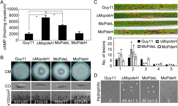
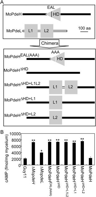
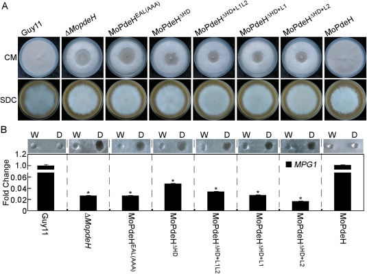
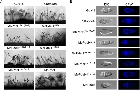
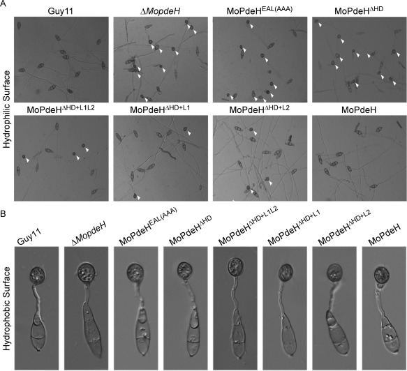
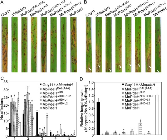
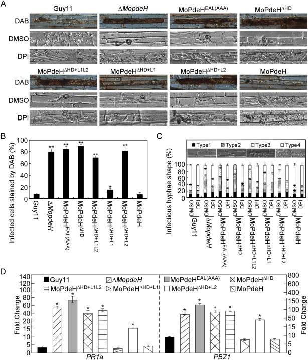
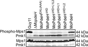
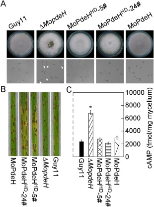
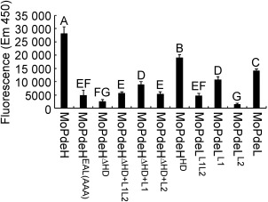
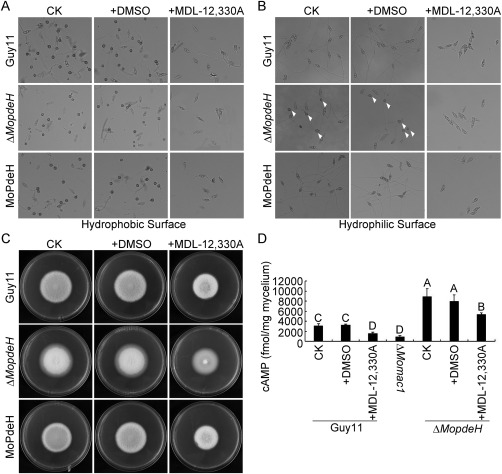
The cyclic adenosine monophosphate (cAMP) signalling pathway mediates signal communication and sensing during infection-related morphogenesis in eukaryotes. Many studies have implicated cAMP as a critical mediator of appressorium development in the rice blast fungus, Magnaporthe oryzae. The cAMP phosphodiesterases, MoPdeH and MoPdeL, as key regulators of intracellular cAMP levels, play pleiotropic roles in cell wall integrity, cellular morphology, appressorium formation and infectious growth in M. oryzae. Here, we analysed the roles of domains of MoPdeH and MoPdeL separately or in chimeras. The results indicated that the HD and EAL domains of MoPdeH are indispensable for its phosphodiesterase activity and function. Replacement of the MoPdeH HD domain with the L1 and L2 domains of MoPdeL, either singly or together, resulted in decreased cAMP hydrolysis activity of MoPdeH. All of the transformants exhibited phenotypes similar to that of the ΔMopdeH mutant, but also revealed that EAL and L1 play additional roles in conidiation, and that L1 is involved in infectious growth. We further found that the intracellular cAMP level is important for surface signal recognition and hyphal autolysis. The intracellular cAMP level negatively regulates Mps1-MAPK and positively regulates Pmk1-MAPK in the rice blast fungus. Our results provide new information to better understand the cAMP signalling pathway in the development, differentiation and plant infection of the fungus.

Keywords: MoPdeH/L; appressorium formation; cAMP level; pathogenicity; phosphodiesterase activity.


Figures

Figure 1
Figure 1
Phenotypic analysis of the MoPdeL overexpression strain. (A) Measurement of the intracellular cyclic adenosine monophosphate (cAMP) level in the indicated strains. (B) Mycelial autolysis (top panel), conidial morphology (middle panel) and conidiation (bottom panel) of the wild‐type Guy11, ΔMopdeH mutant, MoPdeL (MoPdeL overexpressed in ΔMopdeH mutant) and MoPdeH (ΔMopdeH mutant complemented transformant) strains. CO, conidium. (C) Rice spraying assay with conidial suspensions (5 × 10⁴ spores/mL) prepared from the indicated strains, and lesion type statistical analysis: 0, no lesions; 1, dark‐brown pinpoint lesions; 2, 1.5‐mm brown spots; 3, 2–3‐mm lesions with brown margins; 4, eyespot lesions longer than 3 mm; 5, coalesced lesions infecting 50% or more of the leaf maximum size. Lesions were photographed and measured at 7 days post‐inoculation (dpi). The error bars indicate the standard deviation (SD) of three replicates. Asterisks indicate significant differences at P < 0.01. (D) Appressorium formation on hydrophilic surfaces photographed at 24 h post‐inoculation. White arrowheads indicate appressoria.
Figure 2
Figure 2
Strategy for domain mutation and chimera construction, and intracellular cyclic adenosine monophosphate (cAMP) measurement. (A) MoPdeHEAL(AAA), mutation of EAL to AAA in MoPdeH; MoPdeHΔ HD, deletion of HD domain in MoPdeH; MoPdeHΔ HD+L1L2 , replacement of HD with L1&L2 domain of MoPdeL; MoPdeHΔ HD+L1, replacement of HD with L1 domain of MoPdeL; MoPdeHΔ HD+L2, replacement of HD with L2 domain of MoPdeL. (B) Domain mutation and chimeric transformants, ΔMopdeH mutant and ΔMopdeL mutant, lead to increased accumulation of cAMP levels. Bar chart showing the quantification of intracellular cAMP in the mycelia of the above strains cultured for 2 days in complete medium. Two biological repetitions with three replicates were assayed. The error bars indicate the standard deviation (SD) of three replicates. Asterisks indicate significant differences at P < 0.01.
Figure 3
Figure 3
Defects of the indicated strains in mycelial autolysis and surface hydrophobicity. (A) The indicated strains were inoculated on complete medium (CM) and sporulation medium (SDC) plates for 14 days and photographed. (B) Ten microlitres of water (W) or detergent solution containing 0.02% sodium dodecylsulfate (SDS) and 5 mm ethylenediaminetetraacetic acid (EDTA) (D) were placed on the colony surfaces of the indicated strains cultured on CM plates for 14 days, and photographed after 5 min (top panel); quantitative reverse transcription‐polymerase chain reaction (qRT‐PCR) analysis of the expression of the MPG1 gene in the indicated strains is shown in the bottom panel. Error bars indicate the standard deviation (SD) of three replicates. Asterisks indicate significant differences at P < 0.01.
Figure 4
Figure 4
Conidial production and morphological observation of the indicated strains. (A) Conidial formation was observed under a light microscope after illumination for 14 h on a glass slide under fluorescent light and photographed. (B) Conidia of the wild‐type and transformants were stained with 1 mg/mL calcofluor white (CFW) for 5 min in the dark, and observed under a fluorescence microscope.
Figure 5
Figure 5
Appressorium formation of the indicated strains. (A) Conidia of the strains were incubated on a hydrophilic surface for 24 h and photographed. White arrowheads indicate appressoria. (B) Conidia of the strains were incubated on a hydrophobic surface for 24 h and photographed.
Figure 6
Figure 6
Defects of the indicated strains in pathogenicity. (A) Four millilitres of conidial suspension (5 × 10⁴ spores/mL) of each strain were sprayed onto rice seedlings. Diseased leaves were photographed at 7 days post‐inoculation (dpi). (B) A conidial suspension (1 × 10⁵ spores/mL) was injected into rice sheaths. Diseased leaves were photographed at 5 dpi. White arrowheads indicate injection sites. (C) Lesion type statistical analysis. Lesions were photographed and measured at 7 dpi. (D) The severity of disease was analysed by quantification of Magnaporthe oryzae genomic 28S rDNA relative to rice genomic Rubq1 DNA. Asterisks represent significant difference (P < 0.01).
Figure 7
Figure 7
The indicated strains showed defects in infectious growth and induced a strong host defence response. (A) Conidial suspensions of the indicated strains treated with or without 0.5 µm diphenyleneiodonium (DPI) in dimethylsulfoxide (DMSO) were injected in rice leaf sheath and incubated for 36 h before staining with 3,3′‐diaminobenzidine (DAB) for 8 h and observed. (B) Percentage of cells with infectious hyphae stained by DAB. Data were analysed from three independent replicates. Asterisks represent significant difference (*P < 0.05, **P < 0.01). (C) Statistical analysis of the type of infectious hyphae treated with DPI or DMSO. DMSO treatment is a negative control. Three independent experiments were replicated. (D) Quantitative reverse transcription‐polymerase chain reaction (qRT‐PCR) analysis of the transcription levels of rice pathogenesis‐related (PR) genes at 48 h post‐inoculation (hpi). The average threshold cycle of triplicate reactions was normalized by the stable expression gene elongation factor 1α (Os03g08020) in Oryza sativa. Experiments were repeated three times with similar results. Asterisks represent significant difference (p < 0.01).
Figure 8
Figure 8
Defects of MoMps1 phosphorylation of the indicated strains. The total protein was harvested from mycelia cultured in liquid complete medium (CM) for 2 days. The phosphorylated MoMps1 was detected by binding of the antiphospho‐p44/42 antibody, with the Mpk1 antibody as a control.
Figure 9
Figure 9
The HD domain rescued the defects of the ΔMopdeH mutant. (A) The indicated strains were inoculated on complete medium (CM) for 14 days and photographed (top panel). Conidia of the strains were incubated on a hydrophilic surface for 24 h and photographed (bottom panel). White arrowheads indicate appressoria. (B) Rice spraying assay with conidial suspensions (5 × 10⁴ spores/mL) prepared from the indicated strains, and photographed at 7 days post‐inoculation (dpi). (C) Intracellular cyclic adenosine monophosphate (cAMP) level in the mycelia of the indicated strains cultured for 2 days in complete medium and quantified by high‐performance liquid chromatography (HPLC). The error bars indicate standard deviation (SD) of three replicates. Asterisk represents significant difference (P < 0.01).
Figure 10
Figure 10
Detection of protein phosphodiesterase activity in vitro. The target proteins were expressed in Escherichia coli BL21‐CodonPlus (DE3) cells. The phosphate sensor was used to bind free inorganic phosphate (Pi), and fluorescence was read immediately at excitation (420 nm) and emission (450 nm). The error bars indicate the standard deviation of three replicates. Different letters indicate statistically significant differences (P < 0.01).
Figure 11
Figure 11
Suppressed adenylate cyclase in ΔMopdeH rescued its appressorium formation defect on a hydrophilic surface. (A, B) Conidia of the Guy11, ΔMopdeH mutant and complemented strain with 100 μm MDL‐12,330A and equal dimethylsulfoxide (DMSO) as a control were incubated on a hydrophobic or hydrophilic surface for 24 h and photographed. White arrowheads indicate appressoria. (C) Vegetative growth of the indicated strains on complete medium (CM) with or without MDL‐12,330A, and photographed at 7 days post‐inoculation (dpi). (D) Intracellular cAMP level in mycelia treated with or without MDL‐12,330A. The error bars indicate the standard deviation (SD) of three replicates. Different letters indicate statistically significant differences (P < 0.01).

Similar articles

Cited by

References

    1. Arnold, K. , Bordoli, L. , Kopp, J. and Schwede, T. (2006) The SWISS‐MODEL workspace: a web‐based environment for protein structure homology modelling. Bioinformatics, 22, 195–201. - PubMed
    1. Bencina, M. , Panneman, H. , Ruijter, G.J. , Legisa, M. and Visser, J. (1997) Characterization and overexpression of the Aspergillus niger gene encoding the cAMP‐dependent protein kinase catalytic subunit. Microbiology, 143, 1211–1220. - PubMed
    1. Benkert, P. , Biasini, M. and Schwede, T. (2011) Toward the estimation of the absolute quality of individual protein structure models. Bioinformatics, 27, 343–350. - PMC - PubMed
    1. Biasini, M. , Bienert, S. , Waterhouse, A. , Arnold, K. , Studer, G. , Schmidt, T. , Kiefer, F. , Gallo Cassarino, T. , Bertoni, M. , Bordoli, L. and Schwede, T. (2014) SWISS‐MODEL: modelling protein tertiary and quaternary structure using evolutionary information. Nucleic Acids Res. 42, W252–W258. - PMC - PubMed
    1. Bobrov, A.G. , Kirillina, O. and Perry, R.D. (2005) The phosphodiesterase activity of the HmsP EAL domain is required for negative regulation of biofilm formation in Yersinia pestis . FEMS Microbiol. Lett. 247, 123–130. - PubMed

Publication types

MeSH terms

LinkOut - more resources