. 2017 Dec;24(12):2066-2076.
doi: 10.1038/cdd.2017.114. Epub 2017 Jul 28.

ASK1 facilitates tumor metastasis through phosphorylation of an ADP receptor P2Y12 in platelets

Affiliations

ASK1 facilitates tumor metastasis through phosphorylation of an ADP receptor P2Y12 in platelets

Miki Kamiyama et al. Cell Death Differ. 2017 Dec.

Abstract

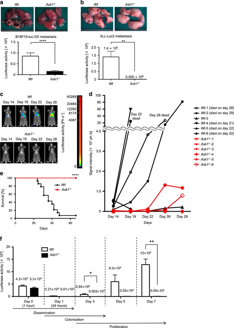
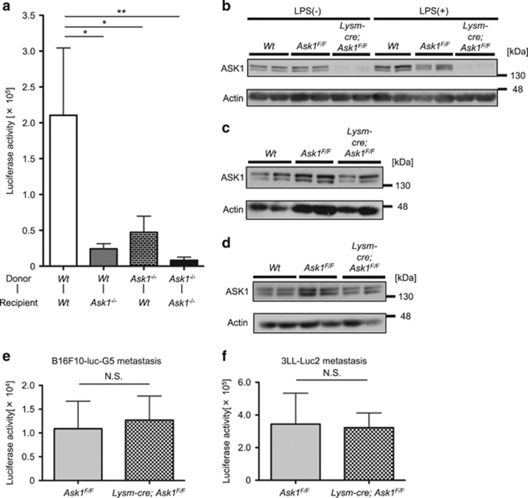
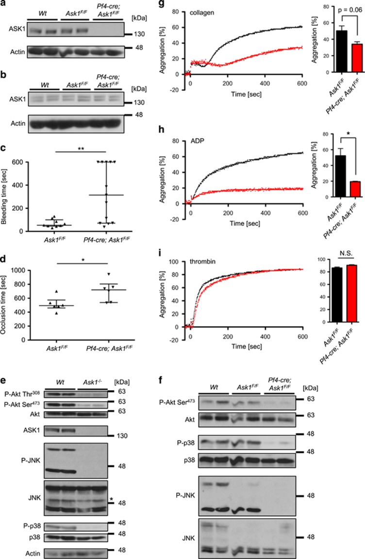
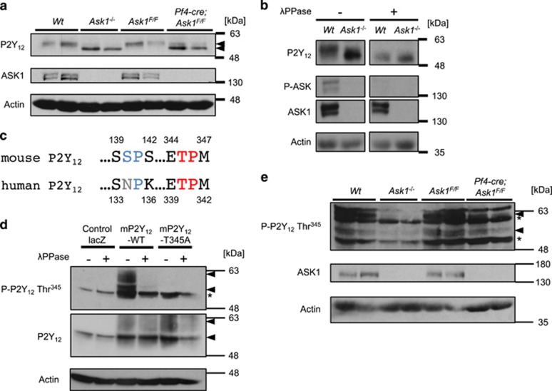
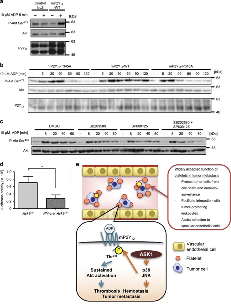
Tumor metastasis is the major cause of deaths in cancer patients and is modulated by intertwined stress-responsive signaling cascades. Here we demonstrate that deletion of stress-responsive apoptosis signal-regulating kinase 1 (Ask1) in platelets results in unstable hemostasis and drastic attenuation of tumor lung metastasis, both of which are attributable to platelet dysfunction. Platelet-specific deletion of Ask1 in mice leads to defects in ADP-dependent platelet aggregation, unstable hemostasis and subsequent attenuation of tumor metastasis. We also revealed that activating phosphorylation of Akt is attenuated in Ask1-deficient platelets, contrary to the previous reports suggesting that Akt is negatively regulated by ASK1. Mechanistically, ASK1-JNK/p38 axis phosphorylates an ADP receptor P2Y12 at Thr345, which is required for the ADP-dependent sustained Akt activity that is vital to normal platelet functions. Our findings offer insight into positive regulation of Akt signaling through P2Y12 phosphorylation as well as MAPK signaling in platelets by ASK1 and suggest that ASK1-JNK/p38 axis provides a new therapeutic opportunity for tumor metastasis.


Conflict of interest statement

The authors declare no conflict of interest.

Figures

Figure 1
Figure 1
Ask1 deficiency in mice attenuates tumor lung metastasis. (a and b) Macroscopic views of metastatic foci in lungs (upper panels) and luciferase activity of lung lysates (lower panels) analyzed 14 days after i.v. injection of B16F10-luc-G5 cells (a) and 3LL-Luc2 cells (b). (a) Wt mice: n=24, Ask1−/− mice: n=23. (b) Wt mice: n=4, Ask1−/− mice: n=4. (c) Mice i.v. injected with 3LL-Luc2 cells were analyzed using an in vivo bioluminescence imaging. (d) Luciferase signal intensity from 3LL-Luc2 cells in the lungs was measured over time using an in vivo bioluminescence imaging (Wt mice: n=6, Ask1−/− mice: n=6). Unless otherwise indicated, the mice survived until the end of the experiment (day 29). (e) The survival of mice i.v. injected with 3LL-Luc2 cells was monitored (Wt mice: n=14, Ask1−/− mice: n=15). (f) Luciferase activity of the lung lysates at indicated time points after tail vein injection of 3LL-Luc2 cells (n=3 per group). The data are shown as the mean±s.e.m. ****P<0.0001; **P<0.01; *P<0.05. Unpaired Student’s t-tests (a, b and f) and Gehan–Breslow–Wilcoxon test (e)
Figure 2
Figure 2
Ask1 deficiency in multiple cell types other than myeloblasts attenuates tumor lung metastasis. (a) Luciferase activity of lung lysates of bone marrow chimeric mice 7 days after i.v. injection of 3LL-Luc2 cells (Wt-Wt mice: n=14, Wt-Ask1−/− mice: n=12, Ask1−/−-Wt mice: n=14, Ask1−/−-Ask1−/− mice: n=16). (bd) Western blot of peritoneal macrophages (b), platelets (c) and lungs (d) of Wt, Ask1F/F and Lysm-cre; Ask1F/F mice (n=2 each). (e and f) Luciferase activity of lung lysates 14 days after i.v. injection of B16F10-luc-G5 cells (e) and 3LL-Luc2 cells (f). (e) Ask1F/F mice: n=7, Lysm-cre; Ask1F/F mice: n=8, (f) Ask1F/F mice: n=10, Lysm-cre; Ask1F/F mice: n=14. The data are shown as the mean±s.e.m. **P<0.01; *P<0.05; NS, nonsignificant. One-way ANOVA followed by Ryan’s method (a) and unpaired Student’s t-tests (e and f)
Figure 3
Figure 3
Platelet-specific Ask1 deletion leads to unstable hemostasis and attenuation in Akt activity. (a and b) Western blot of platelets (a) and lungs (b) of Wt, Ask1F/F and Pf4-cre; Ask1F/F mice (n=2 each). (c) Bleeding time in the tail bleeding assay (Ask1F/F mice: n=9, Pf4-cre; Ask1F/F mice: n=13). (d) Occlusion time in the femoral artery in the FeCl3-induced thrombosis assay (Ask1F/F mice: n=6, Pf4-cre; Ask1F/F mice: n=6). The data are presented with the median and interquartile range. (e and f) Western blot of platelets of Wt and Ask1−/− mice (e, *non-specific band), and Wt, Ask1F/F and Pf4-cre; Ask1F/F mice (f) (n=2 each). (gi) Platelet aggregation of Ask1F/F and Pf4-cre; Ask1F/F mice induced by 1 μg/ml collagen (g), 10 μM ADP (h) and 0.025 U/ml thrombin (i). Representative traces (left) and the maximal aggregation within 10 min of measurement (right) are presented. The data are presented as the mean±s.e.m. (n=3 each). **P<0.01; *P<0.05; NS, non-significant. Mann–Whitney U-test (c and d) and unpaired Student’s t-tests (gi)
Figure 4
Figure 4
ASK1 regulates the phosphorylation of P2Y12 at Thr345 in platelets. (a) P2Y12 expression in platelets of Wt, Ask1−/−, Ask1F/F and Pf4-cre; Ask1F/F mice (n=2 each). (b) Platelet lysates of Wt and Ask1−/− mice were subjected to buffer or λPPase to examine the impact of dephosphorylation on the bands for P2Y12. (c) Intracellular consensus phosphorylation sites for JNK and p38 ((S/T)P sites) in mouse P2Y12 were compared with those of human P2Y12. The SP site of mouse P2Y12 denoted in blue is not conserved in human P2Y12. (d) Evaluation of the specificity of the phospho-P2Y12 Thr345 antibody. CHO cells exogenously expressing mP2Y12-WT, T345A or control lacZ were stimulated with 10 μM ADP for 5 min, lysed and subjected to dephosphorylation by λPPase. (e) Western blot of mouse platelets of Wt, Ask1−/−, Ask1F/F and Pf4-cre; Ask1F/F mice (n=2 each) (arrowhead; P-P2Y12 Thr345 or P2Y12, *nonspecific band)
Figure 5
Figure 5
ASK1-JNK/p38 axis regulates sustained Akt activation induced by ADP stimulation and platelet-dependent tumor metastasis. (a) Akt phosphorylation by ADP stimulation in CHO cells exogenously expressing control lacZ or mouse P2Y12 (mP2Y12)-WT. (b) The phosphorylation-defective (T345A) and recycling-defective (P346A) mutants of mP2Y12 lost sustained Akt signaling by ADP stimulation in CHO cells. (c) JNK and p38 inhibition synergistically suppressed sustained Akt signaling induced by ADP stimulation in CHO cells exogenously expressing mP2Y12-WT. (d) Luciferase activity of lung lysates 14 days after i.v. injection of B16F10-luc-G5 cells (Ask1F/F mice: n=11, Pf4-cre; Ask1F/F mice: n=10). The data are presented as the mean±s.e.m. *P<0.05. Unpaired Student’s t-tests (d). (e) ASK1-JNK/p38 axis in platelets facilitates ADP-induced Akt activity by phosphorylation of mP2Y12 at Thr345 and platelet-dependent tumor metastasis

Comment in

References

    1. Chaffer CL, Weinberg RA. A perspective on cancer cell metastasis. Science 2011; 331: 1559–1564. - PubMed
    1. Widmann C, Gibson S, Jarpe MB, Johnson GL. Mitogen-activated protein kinase: conservation of a three-kinase module from yeast to human. Physiol Rev 1999; 79: 143–180. - PubMed
    1. Murakami S, Noguchi T, Takeda K, Ichijo H. Stress signaling in cancer. Cancer Sci 2007; 98: 1521–1527. - PMC - PubMed
    1. Wagner EF, Nebreda ÁR. Signal integration by JNK and p38 MAPK pathways in cancer development. Nat Rev Cancer 2009; 9: 537–549. - PubMed
    1. Ichijo H, Nishida E, Irie K, Dijke P, Saitoh M, Moriguchi T et al. Induction of apoptosis by ASK1, a mammalian MAPKKK that activates SAPK/JNK and p38 signaling pathways. Science 1997; 275: 90–94. - PubMed

MeSH terms