Feasibility of Dose-reduced Chest CT with Photon-counting Detectors: Initial Results in Humans
- PMID: 28753389
- PMCID: PMC5708286
- DOI: 10.1148/radiol.2017162587
Feasibility of Dose-reduced Chest CT with Photon-counting Detectors: Initial Results in Humans
Abstract
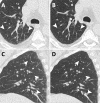
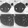
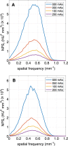
Purpose To investigate whether photon-counting detector (PCD) technology can improve dose-reduced chest computed tomography (CT) image quality compared with that attained with conventional energy-integrating detector (EID) technology in vivo. Materials and Methods This was a HIPAA-compliant institutional review board-approved study, with informed consent from patients. Dose-reduced spiral unenhanced lung EID and PCD CT examinations were performed in 30 asymptomatic volunteers in accordance with manufacturer-recommended guidelines for CT lung cancer screening (120-kVp tube voltage, 20-mAs reference tube current-time product for both detectors). Quantitative analysis of images included measurement of mean attenuation, noise power spectrum (NPS), and lung nodule contrast-to-noise ratio (CNR). Images were qualitatively analyzed by three radiologists blinded to detector type. Reproducibility was assessed with the intraclass correlation coefficient (ICC). McNemar, paired t, and Wilcoxon signed-rank tests were used to compare image quality. Results Thirty study subjects were evaluated (mean age, 55.0 years ± 8.7 [standard deviation]; 14 men). Of these patients, 10 had a normal body mass index (BMI) (BMI range, 18.5-24.9 kg/m2; group 1), 10 were overweight (BMI range, 25.0-29.9 kg/m2; group 2), and 10 were obese (BMI ≥30.0 kg/m2, group 3). PCD diagnostic quality was higher than EID diagnostic quality (P = .016, P = .016, and P = .013 for readers 1, 2, and 3, respectively), with significantly better NPS and image quality scores for lung, soft tissue, and bone and with fewer beam-hardening artifacts (all P < .001). Image noise was significantly lower for PCD images in all BMI groups (P < .001 for groups 1 and 3, P < .01 for group 2), with higher CNR for lung nodule detection (12.1 ± 1.7 vs 10.0 ± 1.8, P < .001). Inter- and intrareader reproducibility were good (all ICC > 0.800). Conclusion Initial human experience with dose-reduced PCD chest CT demonstrated lower image noise compared with conventional EID CT, with better diagnostic quality and lung nodule CNR. © RSNA, 2017 Online supplemental material is available for this article.
Figures


References
-
- Guerra P, Santos A, Darambara DG. Development of a simplified simulation model for performance characterization of a pixellated CdZnTe multimodality imaging system. Phys Med Biol 2008;53(4):1099–1113. - PubMed
-
- Tanguay J, Kim HK, Cunningham IA. The role of x-ray Swank factor in energy-resolving photon-counting imaging. Med Phys 2010;37(12):6205–6211. - PubMed
-
- Weidinger T, Buzug TM, Flohr T, et al. Investigation of ultra low-dose scans in the context of quantum-counting clinical CT. In: Pelc NJ, Nishikawa RM, Whiting BR, eds. Proceedings of SPIE: medical imaging 2012—physics of medical imaging. Vol 8313. Bellingham, Wash: International Society for Optics and Photonics, 2012; 83134B.
-
- Symons R, Cork TE, Lakshmanan MN, et al. Dual-contrast agent photon-counting computed tomography of the heart: initial experience. Int J Cardiovasc Imaging 2017 Mar 13. [Epub ahead of print] - PubMed
Publication types
MeSH terms
Grants and funding
LinkOut - more resources
Full Text Sources
Other Literature Sources
Medical
Molecular Biology Databases

