Review
. 2017 Jul 27;170(3):414-427.
doi: 10.1016/j.cell.2017.07.009.

How Ligands Illuminate GPCR Molecular Pharmacology

Affiliations
Review

How Ligands Illuminate GPCR Molecular Pharmacology

Daniel Wacker et al. Cell. .

Abstract

G protein-coupled receptors (GPCRs), which are modulated by a variety of endogenous and synthetic ligands, represent the largest family of druggable targets in the human genome. Recent structural and molecular studies have both transformed and expanded classical concepts of receptor pharmacology and have begun to illuminate the distinct mechanisms by which structurally, chemically, and functionally diverse ligands modulate GPCR function. These molecular insights into ligand engagement and action have enabled new computational methods and accelerated the discovery of novel ligands and tool compounds, especially for understudied and orphan GPCRs. These advances promise to streamline the development of GPCR-targeted medications.


Figures

Figure 1
Figure 1. Different ligand-stabilized GPCR conformations cause binding and activation of distinct signal transducers including G proteins and arrestins
(Left) Crystal structure of β2AR (light blue cartoon) coupled to Gαs (blue) Gβ (orange) Gγ (green) heterotrimer (PDB ID: 3SN6 (Rasmussen et al., 2011b)) illustrates G protein-mediated signaling. Upon heterotrimer activation, subunits dissociate and Gα modulates second messenger production such as cAMP production through Gαs-mediated activation of adenylyl cyclase. Gβγ modulates separate Gα-independent downstream signaling networks such as ion channels, phospholipases, and receptor kinases. (Right) Crystal structure of rhodopsin (light blue cartoon) coupled to β-arrestin (salmon) (PDB ID: 4ZWJ (Kang et al., 2015)) illustrates arrestin mediated effects such as receptor internalization or activation of kinase signaling networks.
Figure 2
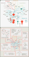
Figure 2. GPCRome wide targets of approved and marketed medications and how ligands uncover unknown GPCR physiology towards potential therapeutic applications
(A) Sphere size corresponds to number of approved drugs for highlighted therapeutic GPCR target with antagonists, agonists, and negative allosteric modulators shown in red, green, and blue, respectively. Phylogenetic tree of the GPCRome highlights the small fraction of GPCRs that are currently targeted by approved medications. (B) Representative examples and their structures are shown for compounds used to identify previously unknown pharmacology at various receptors. Phylogenetic tree of the GPCRome highlights the diversity of GPCRs identified as off-targets.
Figure 3
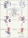
Figure 3. Molecular and structural pharmacology extend the ternary complex model for quantitative description of drug action at GPCRs
Several ligand bound inactive receptor states (R’L, R”L …) and ligand bound active receptor states (R*1L, R*2L …), as well as ternary complex structures of ligand and effector bound active receptor states (R*GL, R*AL) (PDB ID: 3SN6 (Rasmussen et al., 2011b), PDB ID: 4ZWJ (Kang et al., 2015)) have been structurally characterized by x-ray crystallography. Distinct conformational characteristics such as the sodium binding site of the A2AAR (PDB ID: 4EIY (Liu et al., 2012b)), the ionic lock of a nanobody stabilized β2AR (PDB ID: 5JQH (Staus et al., 2016)), the PIF motif of the 5-HT2B receptor (PDB ID: 4IB4 (Wacker et al., 2013), PDB ID: 3NY8 (Wacker et al., 2010)), and the NPxxY motif of a nanobody bound β2AR (PDB ID: 3NY8 (Wacker et al., 2010), PDB ID: 3P0G (Rasmussen et al., 2011a)), highlight diverse GPCR activation states.
Figure 4
Figure 4. Crystal structures of different GPCR-ligand complexes highlight the diverse locations of ligand binding sites
Ligands are shown as stick models with transparent surfaces, receptors are shown in cartoon representation in light blue. Complexes show retinal-bound RHO, LY2119620-bound CHRM2, sodium-bound ADORA2A, ergotamine-bound HTR2B, CCR2-RA-[R]-bound CCR2, MK-0893-bound GCGR, CP-376395-bound CRHR1, and BPTU-bound P2RY1.
Figure 5
Figure 5. Computational approaches generate novel tool compounds for GPCRs
As shown here, the “Pickpocketing” approach identifies GPCRs with potentially related ligand binding properties according to ligand contact strength informed pocket alignment. Similar ligand binding properties between the Neuropeptide S receptor (NPS) and the orphan GPCR GPR37L1 identified the NPS receptor ligand SHA68 as a novel ligand to interrogate GPR37L1 function. The figure is extensively modified from Ngo et al (2017) with permission.
Figure 6
Figure 6

Similar articles

Cited by

References

    1. Airan RD, Thompson KR, Fenno LE, Bernstein H, Deisseroth K. Temporally precise in vivo control of intracellular signalling. Nature. 2009;458:1025–1029. - PubMed
    1. Aitken M, Berndt ER, Cutler DM. Prescription drug spending trends in the United States: looking beyond the turning point. Health Aff (Millwood) 2009;28:w151–160. - PubMed
    1. Alexander SP, Davenport AP, Kelly E, Marrion N, Peters JA, Benson HE, Faccenda E, Pawson AJ, Sharman JL, Southan C, Davies JA, Collaborators, C. The Concise Guide to PHARMACOLOGY 2015/16: G protein-coupled receptors. Br J Pharmacol. 2015;172:5744–5869. - PMC - PubMed
    1. Allen JA, Roth BL. Strategies to discover unexpected targets for drugs active at G protein-coupled receptors. Annu Rev Pharmacol Toxicol. 2011a;51:117–144. - PubMed
    1. Allen JA, Yost JM, Setola V, Chen X, Sassano MF, Chen M, Peterson S, Yadav PN, Huang XP, Feng B, Jensen NH, Che X, Bai X, Frye SV, Wetsel WC, Caron MG, Javitch JA, Roth BL, Jin J. Discovery of beta-arrestin-biased dopamine D2 ligands for probing signal transduction pathways essential for antipsychotic efficacy. Proc Natl Acad Sci U S A. 2011b;108:18488–18493. - PMC - PubMed