. 2017 Jul 28;13(7):e1006913.
doi: 10.1371/journal.pgen.1006913. eCollection 2017 Jul.

The RhoJ-BAD signaling network: An Achilles' heel for BRAF mutant melanomas

Affiliations

The RhoJ-BAD signaling network: An Achilles' heel for BRAF mutant melanomas

Rolando Ruiz et al. PLoS Genet. .

Abstract

Genes and pathways that allow cells to cope with oncogene-induced stress represent selective cancer therapeutic targets that remain largely undiscovered. In this study, we identify a RhoJ signaling pathway that is a selective therapeutic target for BRAF mutant cells. RhoJ deletion in BRAF mutant melanocytes modulates the expression of the pro-apoptotic protein BAD as well as genes involved in cellular metabolism, impairing nevus formation, cellular transformation, and metastasis. Short-term treatment of nascent melanoma tumors with PAK inhibitors that block RhoJ signaling halts the growth of BRAF mutant melanoma tumors in vivo and induces apoptosis in melanoma cells in vitro via a BAD-dependent mechanism. As up to 50% of BRAF mutant human melanomas express high levels of RhoJ, these studies nominate the RhoJ-BAD signaling network as a therapeutic vulnerability for fledgling BRAF mutant human tumors.


Conflict of interest statement

The authors have declared that no competing interests exist.

Figures

Fig 1
Fig 1. RhoJ regulates melanoma tumor development.
(A) Loss of RhoJ prolongs survival of BRAFV600E and PTEN null tumors. Kaplan-Meier survival analysis of 4-OHT treated BrafCA/+; Ptenfl/fl; Tyr::CreERT2; RhoJ+/+ (n = 12) and BrafCA/+; Ptenfl/fl;Tyr::CreERT2; RhoJ-/- (n = 12) mice. Log-rank test demonstrates significant difference between BrafCA/+; Ptenfl/fl;Tyr::CreERT2; RhoJ+/+ and BrafCA/+; Ptenfl/fl;Tyr::CreERT2; RhoJ-/- (p<0.0001). (B) RhoJ deletion prolongs the survival of mice carrying BRAFV600E and PTEN haploinsufficient tumors. Kaplan-Meier survival curves of 4-OHT treated BrafCA/+; Ptenfl/+; Tyr::CreERT2; RhoJ+/+ (n = 19), and BrafCA/+; Ptenfl/+; Tyr::CreERT2; RhoJ-/- (n = 13) mice. Log-rank test demonstrates significant difference between BrafCA/+; Ptenfl/+; Tyr::CreERT2; RhoJ+/+ and BrafCA/+; Ptenfl/+; Tyr::CreERT2; RhoJ-/- (p<0.0001). (C) RhoJ deletion reduces lung metastasis. Lung metastases were compared in aged-matched (P30) mice using a dissection microscope (**p = 0.003, n = 8, Student’s t test). Error bars represent standard error of mean (SEM). (D) RhoJ deletion inhibits melanoma development. The amount of pigment present on the paws at day 17 was analyzed with ImageJ (*p = 0.04, n = 5, T-test). Error bars represent SEM. (E) RhoJ deletion delays melanoma progression. Paws of 4-OHT treated mice were imaged at 10, 17, and 24 days post birth.
Fig 2
Fig 2. RhoJ modulates various signaling pathways to promote tumor growth.
(A) Heat map plot of 50 modulated genes upon loss of RhoJ. Hierarchical clustering of RNA-seq normalized read counts ranging from less frequently expressed (dark blue) to overexpressed (dark red) genes. (B) RhoJ KO mice have a greater number of white hairs than RhoJ WT mice. Images of 8-month old mice show that loss of RhoJ induces accumulation of white hairs. (C) The number of white hairs over an area of 1 in2 were counted and quantified using ImageJ (**p = 0.002, Student’s t-test). Error bars indicate SEM. (D) Loss of RhoJ results in fewer PMEL17+ follicular melanocyte stem cells. Double labeling for the melanosome protein PMEL17 (red) in hair follicle melanocyte stem cells (KIT+, green) at telogen demonstrates a noticeable reduction in the intensity and extent of PMEL expression in RhoJ KO mice. White dashed line indicates the extent of the hair follicle. Scale bars: 10μm (E) Quantification of the immunolabeling described in D demonstrates a statistically significant difference between RhoJ WT and RhoJ KO hairs (*p = 0.03, Student’s t test). Error bars indicate standard deviation of the mean. (F) RhoJ regulates oxidative phosphorylation, melanocyte differentiation, and MAP kinase signaling. Based on the differentially expressed genes between melanoma tumors from RhoJWT and RhoJKO mice, Ingenuity Pathway Analysis (IPA) generated an enrichment of canonical pathways regulated by RhoJ based on the literature (pathways potentially regulated by BRAF are highlighted in red). Bars indicate the negative logarithm of the enrichment p-value. The orange dotted line indicated the statistical threshold (p<0.05) for the enrichment canonical pathways. The orange squares indicate the ratio (value of molecules in a given pathway that meet the cutoff criteria, divided by total number of molecules that make up that pathway) for each canonical pathway. (G) FoxC2 is up regulated in mouse tumor samples. Melanoma tumor lysates were prepared for western blot and probed for the indicated Abs. Relative densitometry values are shown below each blot. (H) BAD is up regulated in RhoJ KO mouse tumor samples. Melanoma tumor lysates were prepared for western blot and probed for the indicated Abs. Relative densitometry values are shown below each blot.
Fig 3
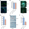
Fig 3. RhoJ regulates the formation of BRAF mutant nevi.
(A) RhoJ deletion does not significantly affect the angiogenesis of BRAF mutant autcthonous melanoma tumors. Tumor sections from age matched (post natal day 30) BRAFV600E and PTEN null RhoJ KO or RhoJ WT mice were stained with smooth muscle actin Ab followed by Alexa-488 secondary Ab to visualize blood vessels. Representative immunofluorescence sections are shown. Scale bars: 100μm. (B) RhoJ KO and RhoJ WT melanomas have similar numbers of vessels. The number of blood vessels in RhoJ WT and RhoJ KO per unit area (412μm x 412 μm) were quantified. The number of blood vessels were not significantly different (p = 0.8, n = 6 (mice) and 3 areas per tumor were counted). (C) RhoJ deletion inhibits nevus formation. MPM images were captured as described in materials and methods from Tyr:CreERT2; BrafV600E; RhoJ+/+ and Tyr:CreERT2; BrafV600E; RhoJ-/-. Colored lines indicate positions being displayed as xy (blue), xz (red) and yz (green) planes. Field of view is 636μm x 636μm Cyan: SHG of collagen; Green: fluorescence of keratin; Yellow and Red–fluorescence of melanin. Nevus indicated by red arrows. Scale bars: 50μm. (D) Nevi were quantified within the upper 50μm from 3-D skin reconstructions. Fewer microscopic nevi could be visualized from the skin surface in RhoJ KO skin as compared to RhoJ wild type skin (p = 0.059, ANOVA, n = 10). (E) RhoJ deletion reduced the number of macroscopic nevi visualized from the skin surface. Skin samples were fixed in 10% formalin for 36 hours and dehydrated in a series of increasing alcohol concentrations, and nevi were visualized by a dissecting microscope. Red arrows indicate pigmentation that could be visualized on the skin surface. (F) Macroscopic nevi were quantified on skin samples that measured 25mm2. RhoJ KO samples contained fewer nevi (p = 0.07, Student’s t-test, n = 4).
Fig 4
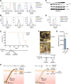
Fig 4. RhoJ signals through PAK1 and is expressed in a subpopulation of Braf mutant human tumors.
(A) RhoJ is expressed in melanoma cell lines containing BRAFV600E mts. Lysates from various melanoma cell lines containing WT BRAF or BRAFV600E mt were prepared and immunoblotted with the indicated Abs. BRAF and PTEN status are indicated below each cell line. (B) RhoJ is activated in melanoma cells. Melanoma cell lysates were analyzed in their initial state (“prior to unloading” lane) or after exchange and reloading with either GDP or GTPγS. Protein lysates were then incubated with PAK-PBD Agarose beads and precipitated. Precipitated lysates were subjected to immunoblotting using the indicated Abs. (C) RhoJ is expressed higher in stage III melanoma tumors. Tumors were scored for RhoJ expression and quantified using standard H-score criteria (**p<0.001). Stage IV tumors are a mixture of primary tumors and metastatic tumors. (D) RhoJ expression is higher in BRAFV600E melanoma clinical specimens. TCGA SKCM RNA-seq data for RhoJ expression was compared between BRAFWT (n = 168) and BRAFV600E (n = 130) melanoma specimens (*p = 0.02, Student's t-test). (E) BRAFV600E and RhoJ status among AJCC stage III and stage IV melanoma tumors. Tissue microarrays were stained with an anti-BRAF mutant specific anti-S100, and an anti-RhoJ Abs as indicated. The percentage of tumors that stained with S100 (positive control) Ab and also stained with RhoJ and/or the mutant BRAF Ab was calculated.
Fig 5
Fig 5. PAK inhibition induces apoptosis via BAD and blocks the progression of BRAF mutant melanomas.
(A) FRAX597 inhibits PAK1 activation and induces apoptosis in BRAFV600E melanoma cell lines. Apoptosis was quantified with annexin-V labeling and flow cytometry. (B) FRAX597 does not induce apoptosis in cell lines that do not express detectable RhoJ. Flow cytometry analysis of melanoma cell lines treated with vehicle, vemurafenib (5μM), FRAX597 (1μM), or trametinib (0.1μM). (C) PAK inhibition decreases the phosphorylation of BADSer 136. Melanoma cell line WM3248 harboring the BRAFV600E mutation was treated with an increasing concentration of FRAX597 (0μM, 0.2μM, 0.5μM, 1μM, 2.5μM, 5μM) and immunoblotted with the indicated antibodies. (D) Over expression of pcDNA3 BADS136E phosphomimetic rescued survival after FRAX597 treatment. Cells were transfected with Lipofectamine 3000 according to the manufacturer’s instructions and treated with FRAX or DMSO. Apoptosis was measured with flow cytometry. (E) PAK inhibition prolongs survival in BrafV600E PTEN null mice. Melanoma was induced in adult mice at P21, P22, and P23 with topical treatment of 4-OHT (25mg/mL) as previously described (18). Vehicle or FRAX597 (100mg/kg P.O. Q.D.) was administered for six days between P28 and P33. Mice were euthanized when the tumor burden exceeded the minimum restrictions as advised by veterinary technicians, and Kaplan-Meier survival curves were generated for BrafCA/+; Ptenfl/fl; Tyr::CreERT2; RhoJ+/+ vehicle (n = 5) or FRAX597 (n = 5) treated animals. Log-rank test demonstrates significant difference between the two groups (**p<0.002, Log-rank test). (F) PAK inhibition delays tumor formation. Mice were treated with 4-OHT at P21, P22, and P23 on their backs to induce melanoma. Tumors were allowed to progress for 2 weeks prior to drug treatment. Mice were either administered FRAX597 or Vehicle for six days. Mice were euthanized after six days of drug treatment and images were taken with a dissecting microscope. Tumors were visible in vehicle treated mice (arrows) but smaller lesions were visible FRAX597 treated mice (arrowheads). (G) Disruption of RhoJ-PAK signaling blocks the formation of nevi and the growth of melanoma. The area of pigmented lesions (developing melanomas) from F was quantified (A.U. arbitrary units) and analyzed (*p = 0.04, n = 3, Student t-test). Error bars indicate SEM. (H) RhoJ expression enables melanocytes to differentiate and migrate out of the hair follicle during the process of nevogenesis and evolve into melanoma tumors. Perturbation of RhoJ-Pak signaling prevents melanocytes from differentiating normally, which leads to loss of pigment in hair, impairment of melanocytes to migrate out of the hair to form nevi, and prevent transformation into melanoma as efficiently.

Similar articles

Cited by

References

    1. Campisi J. Suppressing cancer: the importance of being senescent. Science. 2005;309(5736):886–7. Epub 2005/08/06. doi: 10.1126/science.1116801 . - DOI - PubMed
    1. Michaloglou C, Vredeveld LC, Soengas MS, Denoyelle C, Kuilman T, van der Horst CM, et al. BRAFE600-associated senescence-like cell cycle arrest of human naevi. Nature. 2005;436(7051):720–4. Epub 2005/08/05. doi: 10.1038/nature03890 . - DOI - PubMed
    1. Lowe SW, Cepero E, Evan G. Intrinsic tumour suppression. Nature. 2004;432(7015):307–15. Epub 2004/11/19. doi: 10.1038/nature03098 . - DOI - PubMed
    1. Bild AH, Yao G, Chang JT, Wang Q, Potti A, Chasse D, et al. Oncogenic pathway signatures in human cancers as a guide to targeted therapies. Nature. 2006;439(7074):353–7. Epub 2005/11/08. doi: 10.1038/nature04296 . - DOI - PubMed
    1. Chapman PB, Hauschild A, Robert C, Haanen JB, Ascierto P, Larkin J, et al. Improved survival with vemurafenib in melanoma with BRAF V600E mutation. N Engl J Med. 2011;364(26):2507–16. Epub 2011/06/07. doi: 10.1056/NEJMoa1103782 . - DOI - PMC - PubMed

MeSH terms