. 2017 Jul 28;13(7):e1006937.
doi: 10.1371/journal.pgen.1006937. eCollection 2017 Jul.

The Drosophila Duox maturation factor is a key component of a positive feedback loop that sustains regeneration signaling

Affiliations

The Drosophila Duox maturation factor is a key component of a positive feedback loop that sustains regeneration signaling

Sumbul Jawed Khan et al. PLoS Genet. .

Abstract

Regenerating tissue must initiate the signaling that drives regenerative growth, and sustain that signaling long enough for regeneration to complete. How these key signals are sustained is unclear. To gain a comprehensive view of the changes in gene expression that occur during regeneration, we performed whole-genome mRNAseq of actively regenerating tissue from damaged Drosophila wing imaginal discs. We used genetic tools to ablate the wing primordium to induce regeneration, and carried out transcriptional profiling of the regeneration blastema by fluorescently labeling and sorting the blastema cells, thus identifying differentially expressed genes. Importantly, by using genetic mutants of several of these differentially expressed genes we have confirmed that they have roles in regeneration. Using this approach, we show that high expression of the gene moladietz (mol), which encodes the Duox-maturation factor NIP, is required during regeneration to produce reactive oxygen species (ROS), which in turn sustain JNK signaling during regeneration. We also show that JNK signaling upregulates mol expression, thereby activating a positive feedback signal that ensures the prolonged JNK activation required for regenerative growth. Thus, by whole-genome transcriptional profiling of regenerating tissue we have identified a positive feedback loop that regulates the extent of regenerative growth.


Conflict of interest statement

The authors have declared that no competing interests exist.

Figures
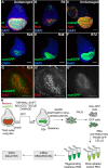
Fig 1
Fig 1. Labeling and isolating regeneration blastema cells.
(A-B) Wing imaginal discs that are undamaged (A) or ablated and at 0 hrs recovery (R0) (B). Green = rnGal4, UAS-EGFP. Red = anti-Nub. Blue = DAPI. (C) Wing imaginal disc showing overlap of anti-Nub immunostaining (red) and expression of the nub-GFP MiMIC enhancer trap (green). (D-F) nub-GFP marks the wing pouch at 24 hrs (D) 48 hrs (E) and 72 hrs (F) after ablation. (G) nub-GFP (green) coincides with the regeneration blastema as defined by a zone of high EdU incorporation (red). (H) Schematic of the mRNA-seq procedure, from tissue ablation through cell dissociation and sort to sequencing and data analysis. Scale bars are100 μm.
Fig 2
Fig 2. Validation of genes identified as upregulated in the regeneration blastema.
Undamaged (A-I) and regenerating (R24) (A’-I’) wing discs. (A-A’) Alp4-lacZ enhancer trap. (B-B’) Atf3-GFP protein trap. (C-C’) chinmo-lacZ enhancer trap. (D-D’) Ets21C-GFP protein trap. (E-E’) mol-lacZ enhancer trap. (F-F’) fru-lacZ enhancer trap. (G-G’) Lamin-GFP protein trap. (H-H’) pigs-GFP enhancer trap. (I-I’) 10xSTAT92E-GFP reporter for STAT activity. Blue dashed line outlines the wing primordium. Scale bars are 100μm.
Fig 3
Fig 3. Validation of genes identified as downregulated in the regeneration blastema.
Undamaged (A-E) and regenerating (R24) (A’-E’) wing discs. (A-A’) dve-lacZ enhancer trap. (B-B’) Hr78-GFP protein trap. (C-C’) NC2β-GFP protein trap. (D-D’) sm-lacZ enhancer trap. (E-E’) Cat-GFP enhancer trap. Blue dashed lines outline the wing primordium. Scale bars are 100 μm.
Fig 4
Fig 4. Genetic assays demonstrating that differentially expressed genes have functional roles in regeneration.
(A) Representative examples of wings from damaged discs that are approximately <25%, 25%, 50%, 75%, and 100% of a normal wing. Scale bar is 1mm. (B-G) Sizes of adult wings after regeneration in control (w1118) and heterozygous mutant animals. Three independent experiments each, error bars are SEM. (B) Sizes of adult wings after regeneration in w1118 and Ets21Cf03639/+ animals. w1118 n = 318 wings, Ets21Cf03639/+ n = 255 wings, p<0.0001 using a chi-squared test. (C) Sizes of adult wings after regeneration in w1118 and CG9336MI03849/+ animals. w1118 n = 374 wings, CG9336MI03849/+ n = 215 wings, p<0.0001 by a chi-squared test. (D) Sizes of adult wings after regeneration in w1118 and Alp407028/+ animals. w1118 n = 239 wings, Alp407028/+ n = 217 wings, p<0.0001 by a chi-squared test. (E) Sizes of adult wings after regeneration in w1118 and Thor06270/+ animals. w1118 n = 224 wings, Thor06270/+ n = 146 wings, p = 0.0021 by a chi-squared test. (F) Sizes of adult wings after regeneration in w1118 and mole02670/+ animals. Three independent experiments, w1118 n = 356 wings, mole02670/+ n = 183 wings, p = 0.00001 by a chi-squared test. (G) Sizes of adult wings after regeneration in w1118, Col4a1k00405/+, and vkgk00236/+ animals. w1118 n = 320 wings, Col4a1k00405/+ n = 71 wings, and vkgk00236/+ n = 134 wings, p<0.0001 by a chi-squared test.
Fig 5
Fig 5. ROS persist in the regeneration blastema and are required for regeneration.
(A-B) DHE staining (red) to detect ROS. The wing pouch is marked with nub-GFP (green). Yellow asterisks mark pockets of cellular debris. (A) Undamaged disc. (B) Regenerating disc at R24. (C-D) Genetic regeneration assays using adult wing size to assess extent of regenerative growth in the imaginal discs. Three independent experiments for each. (C) Sizes of adult wings after regeneration in w1118 and UAS-Sod1/+ animals. w1118 n = 375 wings, UAS-Sod1/+ n = 166 wings, p<0.0001 using a chi-squared test. (D) Sizes of adult wings after regeneration in w1118, UAS-Sod2/+, and UAS-Cat/+ animals. w1118 n = 327 wings, UAS-Sod2/+ n = 332 wings, UAS-Cat/+ n = 361 wings, p<0.0001 using a chi-squared test. (E-H) Anti-Nub marks the wing primordium of w1118 (E,F) and UAS-Sod1 (G,H) regenerating discs at R24 and R48. (I) Quantification of area of the wing primordium as marked by anti-Nub at R24 and R48. w1118 R24 total n = 12 discs, UAS-Sod1 R24 n = 15 discs, w1118 R48 n = 5 discs, UAS-Sod1 R48 n = 10 discs. At R48, p = .0248. (J-K) Pupariation rates. Note that because of the temperature shifts in the ablation protocol the regenerating and undamaged pupariation times cannot be compared to each other. (J) Pupariation timing after regeneration. Three independent experiments, control n = 213 pupae, UAS-Sod1 n = 107 pupae. (K) Pupariation timing during normal development. Three independent experiments, control n = 173 pupae, UAS-Sod1 n = 201 pupae. (L-O) Expression of the TRE-red reporter indicates JNK signaling activity in w1118 (L,M) and UAS-Sod1 (N,O) regenerating discs at R24 and R48. (P) Quantification of TRE-red fluorescence at R24 and R48. *p < .00002. w1118 R24 n = 10 discs, UAS-Sod1 R24 n = 14 discs, w1118 R48 n = 10 discs, UAS-Sod1 R48 n = 14 discs. Scale bars are 100 μm. Error bars are SEM.
Fig 6
Fig 6. moladietz is required for wing disc regeneration.
(A-D) DHE fluorescence (red) indicates the presence of ROS. nub-GFP (green) marks the regenerating wing pouch. (A-B) Confocal slices of a w1118 regenerating disc through the debris field (A,A’) and the disc epithelium (B,B’). Asterisks mark cellular debris in the debris field and in a few folds in the epithelium. Arrow points to the position of the regenerating wing pouch. (C-D) Confocal slices of a mole02670/+ regenerating disc through the debris field (C,C’) and the regenerating epithelium (D,D’). Asterisk and arrow same as above. (E-F) Quantification of DHE fluorescence intensity in the debris fields of w1118 and mole02670/+ regenerating discs (E) and in the regenerating epithelia of w1118 and mole02670/+ regenerating discs and control undamaged discs (F). For R24, three independent experiments, for a total w1118 regenerating n = 12 discs, mole02670/+ regenerating n = 18 discs, w1118 undamaged n = 11 discs. For R48, three independent experiments for a total w1118 regenerating n = 30 discs, mole02670/+ regenerating n = 25 discs, w1118 undamaged n = 10 discs. (G,H) Quantification of GFP fluorescence from a gstD-GFP reporter for ROS-regulated transcription in regenerating w1118 and mole02670/+ discs. For R24, w1118 n = 12 discs, mole02670/+ n = 20 discs. For R48, w1118 n = 12 discs, mole02670/+ n = 10 discs. (I) Adult wing area in w1118 and mole02670/+ male and female wings from undamaged discs and after disc regeneration. Three independent experiments. Undamaged: w1118 females n = 125 wings, w1118 males n = 132 wings, mole02670/+ females n = 82 wings, mole02670/+ males n = 73 wings. Regenerated: w1118 females n = 226 wings, w1118 males n = 134 wings, mole02670/+ females n = 128 wings, mole02670/+ males n = 133 wings. (J-O) Anti-Nub marks the regenerating wing primordium at R0, R24 and R48 in w1118 and mole02670/+ discs. (P) Quantification of the size of the regenerating wing primordium at R0, R24 and R48. R0 w1118 n = 26 and mole02670/+ n = 29, R24 w1118 n = 42 and mole02670/+ n = 41, R48 w1118 n = 29 and mole02670/+ n = 42. Scale bars are 100 μm. Error bars are SEM. **p<0.05, *p<0.005, ***p<0.0002, ****p<0.0001.
Fig 7
Fig 7. NIP is required to sustain JNK signaling during late regeneration.
(A-H) Confocal images of fluorescence from the TRE-red reporter for JNK signaling in w1118 (A-D) and mole02670/+ (E-H) regenerating discs at R0 (A,B), R24 (B,F), R48 (C,G) and R72 (D,H). (I) Quantification of fluorescence intensity of the TRE-red reporter in max projections of the confocal images at R0, because at this time point the epithelium cannot be distinguished from the debris. w1118 n = 10 discs, mole02670/+ n = 14 discs. (J) Quantification of fluorescence intensity of the TRE-red reporter in single slices of the confocal images through the regenerating epithelium at R24, R48, and R72. R24 w1118 n = 11 discs, mole02670/+ n = 11 discs. R48 w1118 = 14 discs, mole02670/+ n = 15 discs. R72 w1118 n = 11 discs, mole02670/+ n = 11 discs. (K) Regeneration assays using adult wing size to assess extent of regenerative growth in the imaginal discs in w1118, mole02670/+, pucE69/+, and mole02670/+;pucE69/+ animals. Two independent experiments, thus error bars are SD. w1118 n = 26 wings, mole02670/+ n = 83 wings, pucE69/+ n = 99 wings, and mole02670/+;pucE69/+ n = 95 wings. p<0.0001 for all comparisons using a chi-squared test. Dashed blue line outlines the wing primordium. Scale bars are 100 μm. Error bars are SEM unless otherwise noted. *p<0.05, **p<0.001, ***p<0.0001.
Fig 8
Fig 8. Expression of mol is regulated by JNK signaling.
(A) Schematic of the mol locus showing the relative positions of three canonical TRE sites. (B,C) Anti-β-galacosidase immunostaining showing expression of the mol-lacZ (green) reporter in control (B) and UAS-JNKDN (C) R24 discs. (D) Quantification of mol-lacZ fluorescence from the immunostaining. w1118 n = 10 discs, UAS-JNKDN n = 10 discs. (E) Anti-β-galacosidase immunostaining showing expression of the mol-lacZ reporter in a hepr75/+ R24 regenerating disc. (F) Quantification of mol-lacZ fluorescence from the immunostaining. w1118 n = 7 discs, hepr75/+ n = 8 discs. Scale bars are 100 μm. Error bars are SEM. **p<0.002.

Similar articles

Cited by

References

    1. Tanaka EM, Reddien PW. The cellular basis for animal regeneration. Dev Cell. 2011. July 19;21(1):172–85. doi: 10.1016/j.devcel.2011.06.016 - DOI - PMC - PubMed
    1. Hobmayer B, Rentzsch F, Kuhn K, Happel CM, Laue von CC, Snyder P, et al. WNT signalling molecules act in axis formation in the diploblastic metazoan Hydra. Nature. 2000. September 14;407(6801):186–9. doi: 10.1038/35025063 - DOI - PubMed
    1. Kawakami Y, Rodriguez Esteban C, Raya M, Kawakami H, Martí M, Dubova I, et al. Wnt/beta-catenin signaling regulates vertebrate limb regeneration. Genes Dev. 2006. December 1;20(23):3232–7. doi: 10.1101/gad.1475106 - DOI - PMC - PubMed
    1. Petersen CP, Reddien PW. Smed-betacatenin-1 is required for anteroposterior blastema polarity in planarian regeneration. Science. 2008. January 18;319(5861):327–30. doi: 10.1126/science.1149943 - DOI - PubMed
    1. Petersen CP, Reddien PW. A wound-induced Wnt expression program controls planarian regeneration polarity. Proc Natl Acad Sci USA. 2009. October 6;106(40):17061–6. doi: 10.1073/pnas.0906823106 - DOI - PMC - PubMed

MeSH terms