. 2017 Jul 28;12(7):e0182001.
doi: 10.1371/journal.pone.0182001. eCollection 2017.

Regeneration of the digestive system in the crinoid Himerometra robustipinna occurs by transdifferentiation of neurosecretory-like cells

Affiliations

Regeneration of the digestive system in the crinoid Himerometra robustipinna occurs by transdifferentiation of neurosecretory-like cells

Nadezhda V Kalacheva et al. PLoS One. .

Abstract

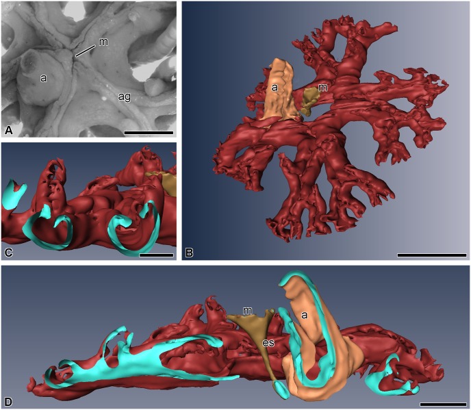
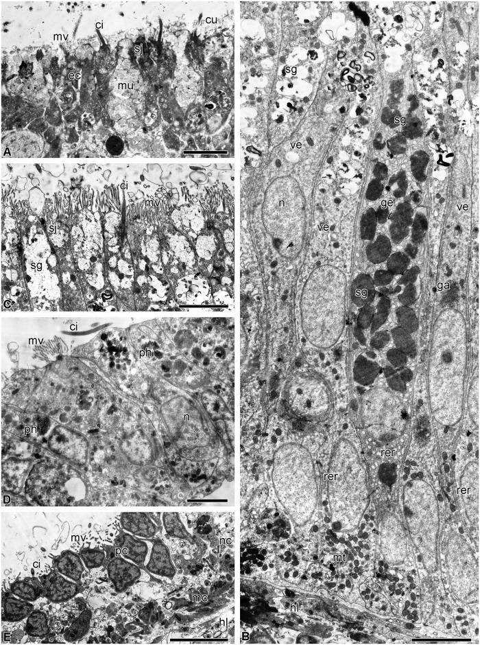
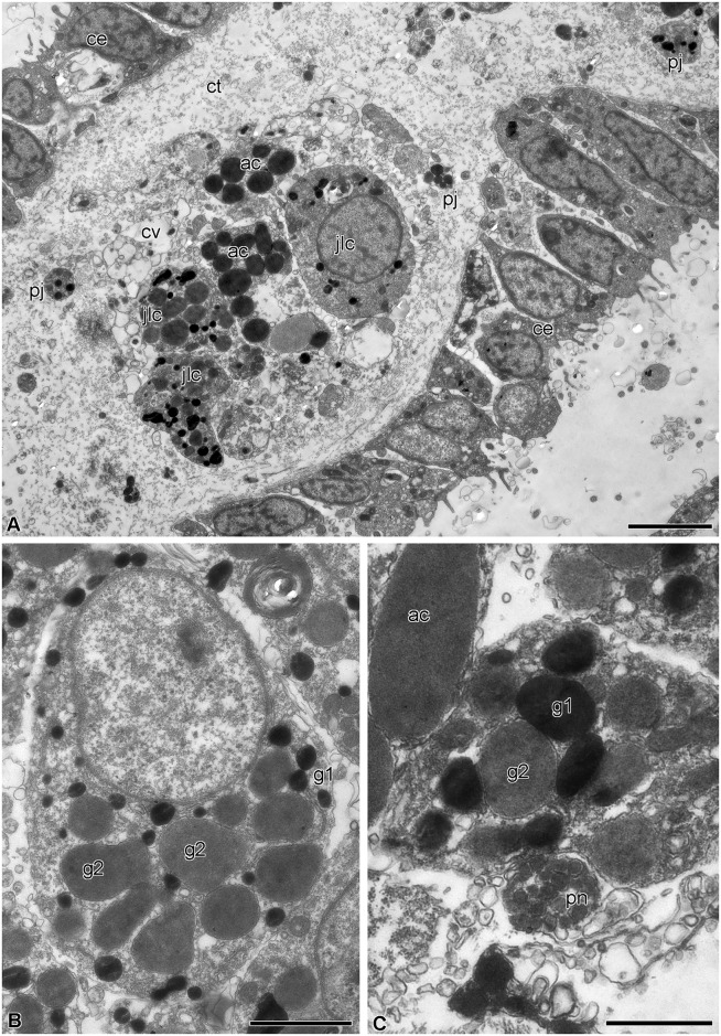
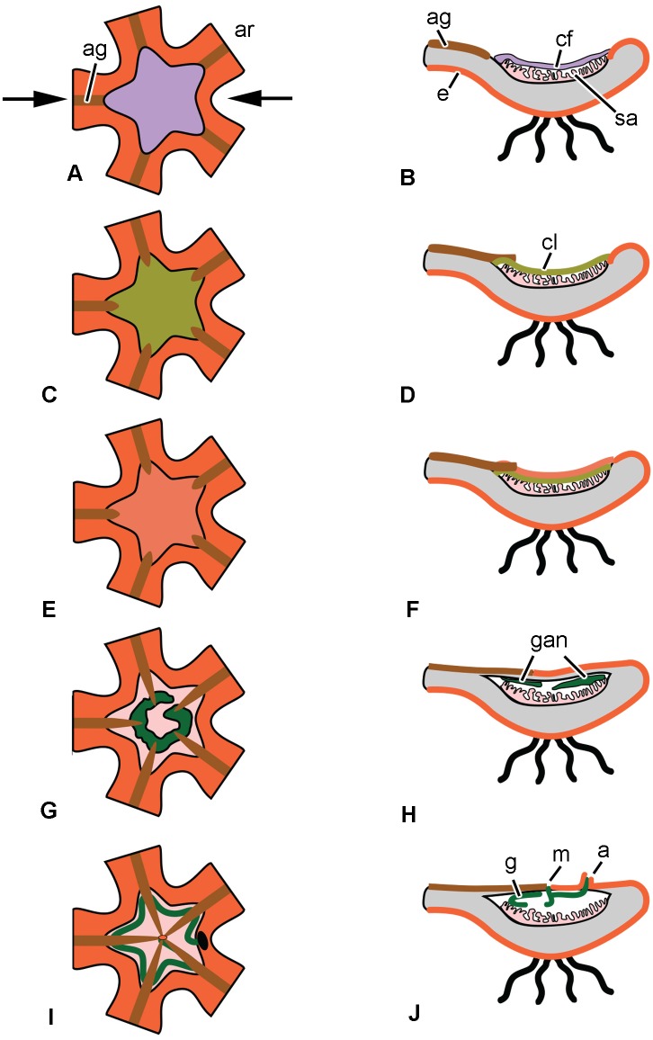
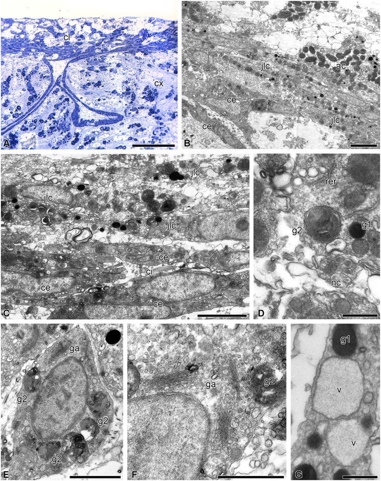
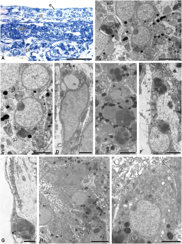
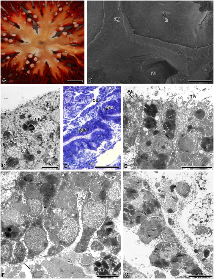
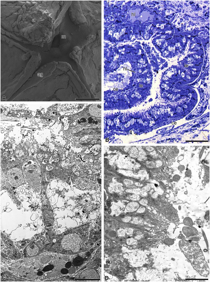
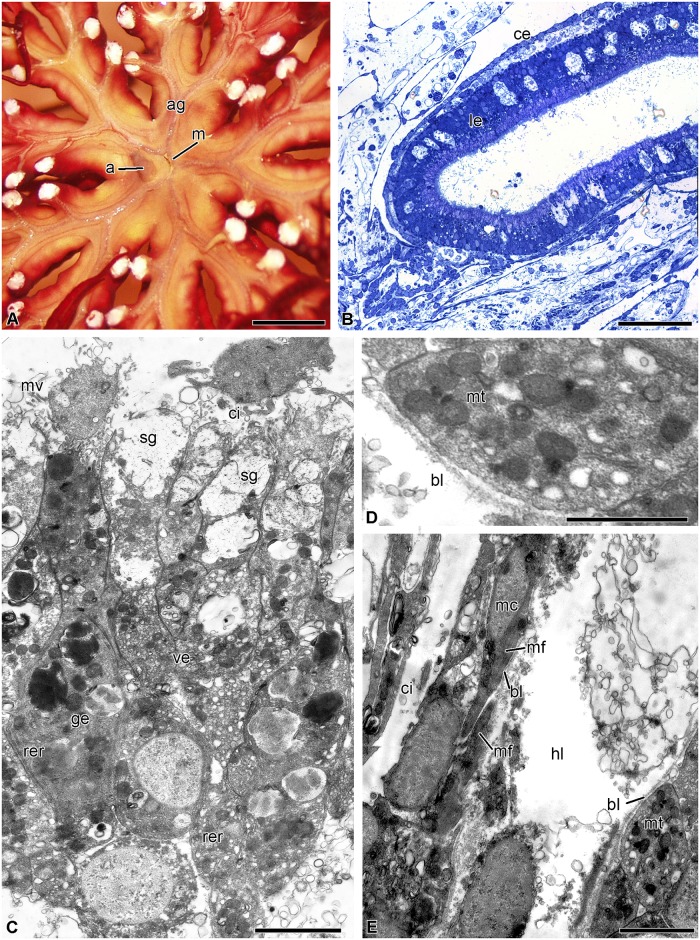
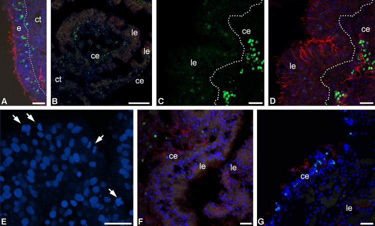
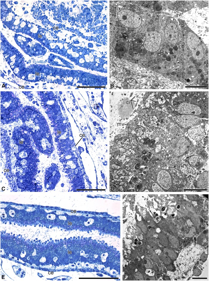
The structure and regeneration of the digestive system in the crinoid Himerometra robustipinna (Carpenter, 1881) were studied. The gut comprises a spiral tube forming radial lateral processes, which gives it a five-lobed shape. The digestive tube consists of three segments: esophagus, intestine, and rectum. The epithelia of these segments have different cell compositions. Regeneration of the gut after autotomy of the visceral mass progresses very rapidly. Within 6 h after autotomy, an aggregation consisting of amoebocytes, coelomic epithelial cells and juxtaligamental cells (neurosecretory neurons) forms on the inner surface of the skeletal calyx. At 12 h post-autotomy, transdifferentiation of the juxtaligamental cells starts. At 24 h post-autotomy these cells undergo a mesenchymal-epithelial-like transition, resulting in the formation of the luminal epithelium of the gut. Specialization of the intestinal epithelial cells begins on day 2 post-autotomy. At this stage animals acquire the mouth and anal opening. On day 4 post-autotomy the height of both the enterocytes and the visceral mass gradually increases. Proliferation does not play any noticeable role in gut regeneration. The immersion of animals in a 10-7 M solution of colchicine neither stopped formation of the lost structures nor caused accumulation of mitoses in tissues. Weakly EdU-labeled nuclei were observed in the gut only on day 2 post-autotomy and were not detected at later regeneration stages. Single mitotically dividing cells were recorded during the same period. It is concluded that juxtaligamental cells play a major role in gut regeneration in H. robustipinna. The main mechanisms of morphogenesis are cell migration and transdifferentiation.


Conflict of interest statement

Competing Interests: The authors have declared that no competing interests exist.

Figures

Fig 1
Fig 1. Three-dimensional reconstruction of the digestive system of H. robustipinna.
(A) General view of the visceral mass (light microscopy). (B) View from oral side. (C) Transverse section (oral-aboral) of radial lateral outgrowth of the gut. (D) Transverse section (oral-aboral) of the gut through central part. a, anal cone; ag, ambulacral groove; es, esophagus; m, mouth; the pale blue color indicates the internal surfaces of the visceral mass in sites of cut. Scale bar: (A and B) 5 mm, (C) 2 mm, (D) 1 mm.
Fig 2
Fig 2. Structure of the luminal epithelium of the digestive tube of H. robustipinna.
(A) Esophagus. (B) General view of the intestinal epithelium. (C) Apical part of the intestinal epithelium. (D) The epithelium of the rectum. (E) Coelomic epithelium of the gut. ci, cilium; cu, cuticle; ec, epitheliocyte; ga, Golgi apparatus; ge, granular enterocyte; hl, hemal lacuna; mu, mucus cell; mc, myoepithelial cell; mt, mitochondria; mv, microvilli; n, nucleus; nc, processes of nerve cells; pc, peritoneocyte; ph, phagosome; rer, rough endoplasmic reticulum; sj, septate junction; sg, secretory granules; ve, vesicular enterocyte. Scale bars: (A) 2 μm, (B-E) 4 μm.
Fig 3
Fig 3. Structure of the septa of the aboral coelom of H. robustipinna.
(A) General view of septa. (B) Juxtaligamental cell. (C) Close apposition of process of juxtaligamental cell and axon (pn). ac, amoebocyte; ce, coelomic epithelium; ct, connective tissue; cv, cell with electron-transparent vacuoles; g1, type 1 granule; g2, type 2 granule; jlc, luxtaligamental cell; pj, process of juxtaligamental cell. Scale bars: (A) 4 μm, (B) 2 μm, (C) 1 μm.
Fig 4
Fig 4. Scheme of the consecutive stages of the regeneration of the digestive system in H. robustipinna.
(A, C, E, G, I)–oral view, (B, D, F, H, J)–transverse section of the calyx (arrows in (A) indicate location of the section plane, (G, I)–epidermis on oral side is not shown). (A, B) Immediately after autotomy. (C, D) 6–12 h post-autotomy. (E, F) 18–24 h post-autotomy. (G, H) 2 days post-autotomy. (I, J) 4 days post-autotomy. a, anal cone; ag, ambulacral groove; ar, arm; cf, coagulated coelomic fluid; cl, cellular layer; e, epidermis; g, gut; gan, gut anlage; m, mouth; sa, septa of the aboral coelom.
Fig 5
Fig 5. Microscopic organization of the cellular layer covering the surface of the calyx at 6–12 h post-autotomy in H. robustipinna.
(A) Transverse (oral-aboral) semithin section of the oral part of the calyx at 6 h post-autotomy. (B) Cellular layer on the surface of the calyx at 6 h post-autotomy. (C) Juxtaligamental cells covering the torn septa of the aboral coelom at 6 h post-autotomy. (D) Transforming type 2 granules at 12 h post-autotomy. (E) Juxtaligamental cell with type 2 granules undergoing destruction at 12 h post-autotomy. (F) Golgi apparatus in the cytoplasm of juxtaligamental cell at 12 h post-autotomy. (G) Large vacuoles in the cytoplasm of juxtaligamental cell at 12 h post-autotomy. ac, amoebocyte; ce, coelomic epithelial cell; ci, cilium; cl, cellular layer; cx, calyx; g1, type 1 granule; g2, type 2 granule; ga, Golgi apparatus; jlc, juxtaligamental cell; nc, process of nerve cell; rer, rough endoplasmic reticulum; v, vacuole. Scale bars: (A) 50 μm, (B) 4 μm, (C, E, F) 2 μm, (D, G) 0.5 μm.
Fig 6
Fig 6. Gut anlage at 18–24 h post-autotomy in H. robustipinna.
(A) Transverse (oral-aboral) semithin section of the cellular layer on the calyx surface at 18 h post-autotomy. (B) Aggregation of juxtaligamental cells and amoebocytes in the gut anlage at 18 h post-autotomy. (C) Juxtaligamental cell with type 2 granules undergoing destruction at 18 h post-autotomy. (D) Polarizing juxtaligamental cell at 18 h post-autotomy. (E) Juxtaligamental cells with amoebocyte granules (*). (F) Cell of coelomic epithelium with amoebocyte granules (*). (G) Epidermal cell with amoebocyte granules (*). (H) Intestinal epithelium composed of transformed juxtaligamental cells at 24 hours post-autotomy. (I) Transformed juxtaligamental cell at 24 h post-autotomy. ac, amoebocyte; cav, cavity; ce, coelomic epithelial cell; ci, cilium; ct, connective tissue; cx, calyx; d, desmosome; e, epidermis; g1, type 1 granule; g2, type 2 granule; gan, gut anlage; jlc, juxtaligamental cell; mt, mitochondrion; sj, septate junction; v, vacuole. Scale bars: (A) 50 μm, (B, H) 4 μm, (C, E-G, I) 2 μm, (D) 1 μm.
Fig 7
Fig 7. Gut anlage at 2 days post-autotomy in H. robustipinna.
(A) General view of the calyx with regenerating visceral mass. (B) Mouth and anal openings. (C) Epidermis with mitotically dividing cell (mi). (D) Gut anlage. (E) Apical part of the enterocytes. (F) Basal part of the enterocytes. (G) Coelomic epithelium of the gut. a, anal opening; ag, ambulacral groove; bl, basal lamina; ce, coelomic epithelial cell; ct, connective tissue; gan, gut anlage; m, mouth; mi, mitosis; mv, microvilli; sg, secretory granules. Scale bars: (A) 5 mm, (B) 400 μm, (D) 50 μm, (C) 5 μm, (E, G) 4 μm, (F) 2 μm.
Fig 8
Fig 8. Digestive system at 4 days post-autotomy in H. robustipinna.
(A) General view of oral surface of the visceral mass. (B) Digestive tube. (C) Luminal epithelium. (D) Apical part of the enterocytes. a, anal cone; ac, amoebocyte; ag, ambulacral groove; ce, coelomic epithelium; ci, cilium; ge, granular enterocyte; hl, hemal lacuna; le, luminal epithelium; m, mouth; mv, microvilli; sg, secretory granule; ve, vesicular enterocyte. Scale bars: (A) 400 μm, (B) 20 μm, (C, D) 4 μm.
Fig 9
Fig 9. Digestive system at 7 days post-autotomy in H. robustipinna.
(A) General view of the oral surface of the visceral mass. (B) Digestive tube. (C) Luminal epithelium. (D) Basal part of the enterocytes. (E) Coelomic epithelium of the gut. a, anal cone; ag, ambulacral groove; bl, basal lamina; ce, coelomic epithelium; ci, cilium; ge, granular enterocyte; hl, hemal lacuna; le, luminal epithelium; m, mouth; mc, myoepithelial cell; mf, myofilaments; mt, mitochondria; mv, microvilli; rer, rough endoplasmic reticulum; sg, secretory granule; ve, vesicular enterocyte. Scale bars (A) 5 mm, (B) 50 μm, (C) 4 μm, (E) 2 μm, (D) 1 μm.
Fig 10
Fig 10. DNA-synthesizing and mitotic cells in the regenerating visceral mass of H. robustipinna.
(A) Epidermis at 2 days post-autotomy; dotted line indicates basal lamina. (B) Transverse section (oral-aboral) of the radial lobe of the visceral mass at 2 days post-autotomy. (C, D) Luminal and coelomic epithelia of the gut at 2 days post-autotomy; C, EdU-labeled nuclei (green channel); D, merged channels. (E) Luminal epithelium with mitoses (arrows) at 2 days post-autotomy. (F) Luminal and coelomic epithelia of the gut at 4 days post-autotomy. (G) Luminal and coelomic epithelia of the gut at 7 days post-autotomy. ce, coelomic epithelium; ct, connective tissue; e, epidermis; le, luminal epithelium; green color, EdU; red color, tubuline; blue color, DAPI. Scale bars: (A, C-E) 20 μm, (B) 100 μm.
Fig 11
Fig 11. Structure of gut of H. robustipinna regenerating 2 days in 10−7 M colchicine solution.
(A, B) The gut of the animals placed in the solution immediately after autotomy. (C, D) The gut of the animals placed in the solution on day 4 post-autotomy. (E, F) The gut of the animals placed in the solution on day 7 post-autotomy. General view of the gut (A, C, E) and fine structure of the luminal epithelium (B, D, F). ce, coelomic epithelium; ci, cilium; hl, hemal lacuna; le, luminal epithelium; mv, microvilli; sg, secretory granules. Scale bars: (A, C, E) 50 μm, (B, D, F) 4 μm.

Similar articles

Cited by

References

    1. Becker SF, Jarriault S (2016) Natural and induced direct reprogramming: mechanisms, concepts and general principles—from the worm to vertebrates. Current Opinion in Genetics & Development 40: 154–163. - PubMed
    1. Eguchi G, Kodama R (1993) Transdifferentiation. Current Opinion in Cell Biology 5: 1023–1028. - PubMed
    1. Eguizabal C, Montserrat N, Veiga A, Belmonte JCI (2013) Dedifferentiation, transdifferentiation, and reprogramming: future irections in regenerative medicine. Seminars in Reproductive Medicine 31: 082–094. - PubMed
    1. Fu L, Zhu X, Yi F, Liu G-H, Belmonte JCI (2014) Regenerative medicine: transdifferentiation in vivo. Cell Research 24: 141–142. doi: 10.1038/cr.2013.165 - DOI - PMC - PubMed
    1. Hu CP, Wu XR, Li QG, Sun ZW, Wang AP, Feng JT, et al. (2013) Proteomic analysis of NGF-induced transdifferentiation of adrenal medullary cells. International Journal of Molecular Medicine 32: 347–354. doi: 10.3892/ijmm.2013.1387 - DOI - PubMed

MeSH terms

LinkOut - more resources