. 2017 Jul 28;13(7):e1006534.
doi: 10.1371/journal.ppat.1006534. eCollection 2017 Jul.

Bacterial effector NleL promotes enterohemorrhagic E. coli-induced attaching and effacing lesions by ubiquitylating and inactivating JNK

Affiliations

Bacterial effector NleL promotes enterohemorrhagic E. coli-induced attaching and effacing lesions by ubiquitylating and inactivating JNK

Xiangpeng Sheng et al. PLoS Pathog. .

Abstract

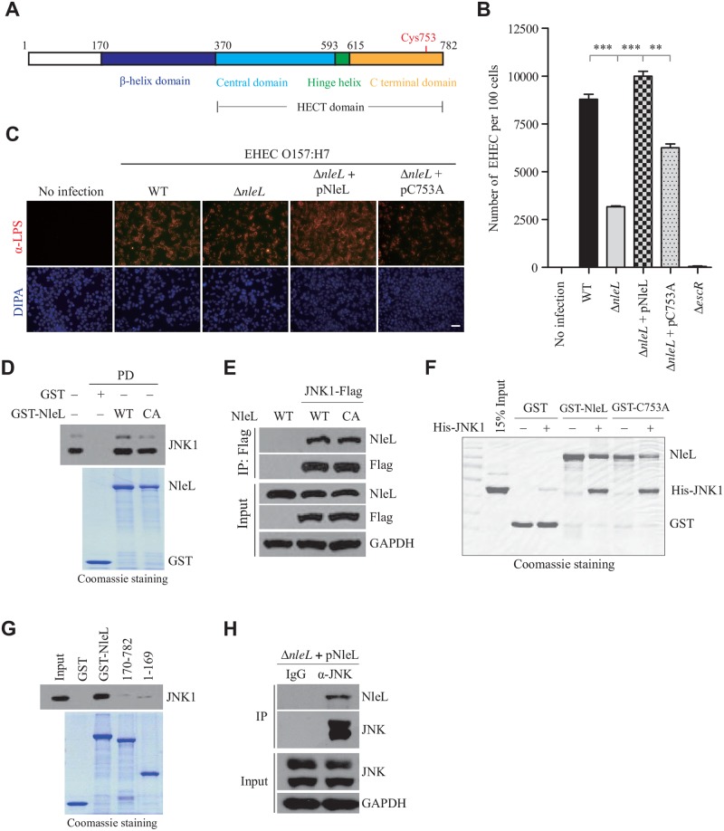
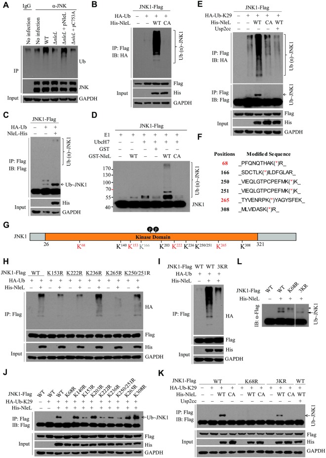
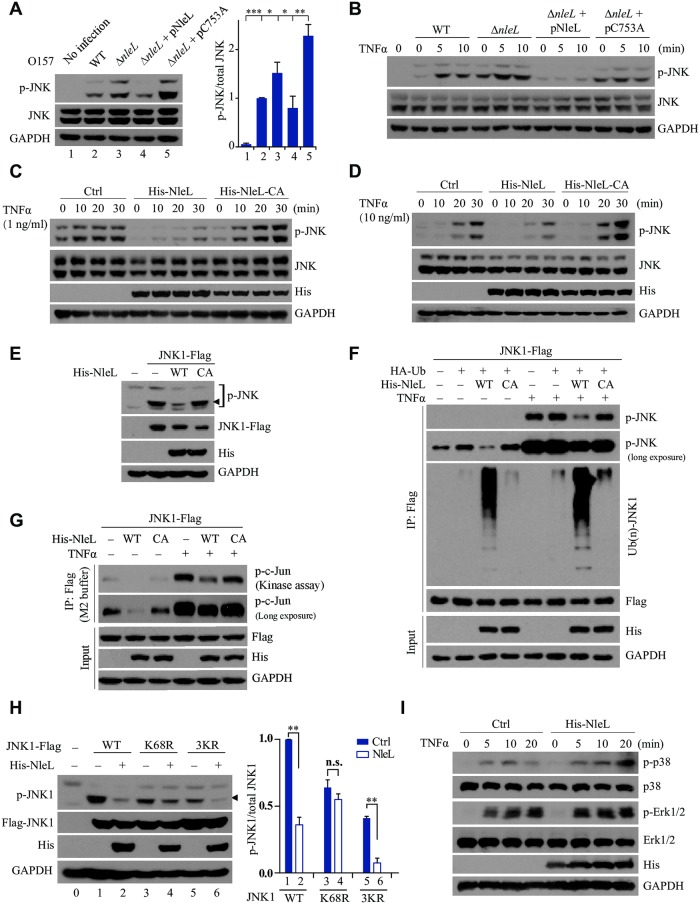
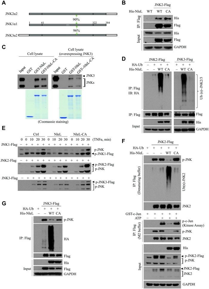
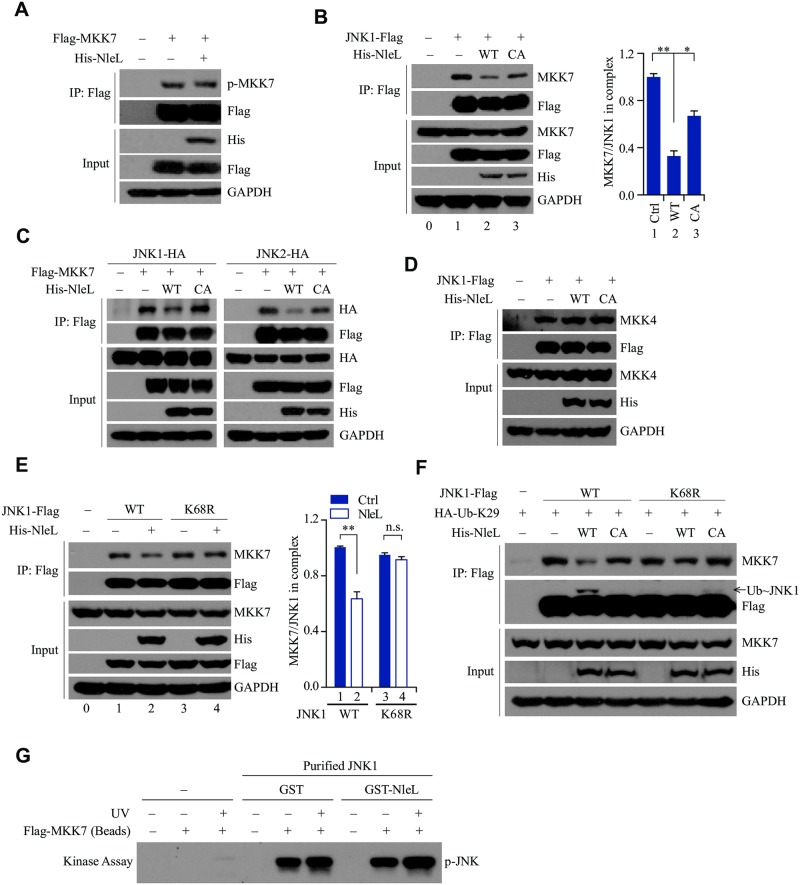
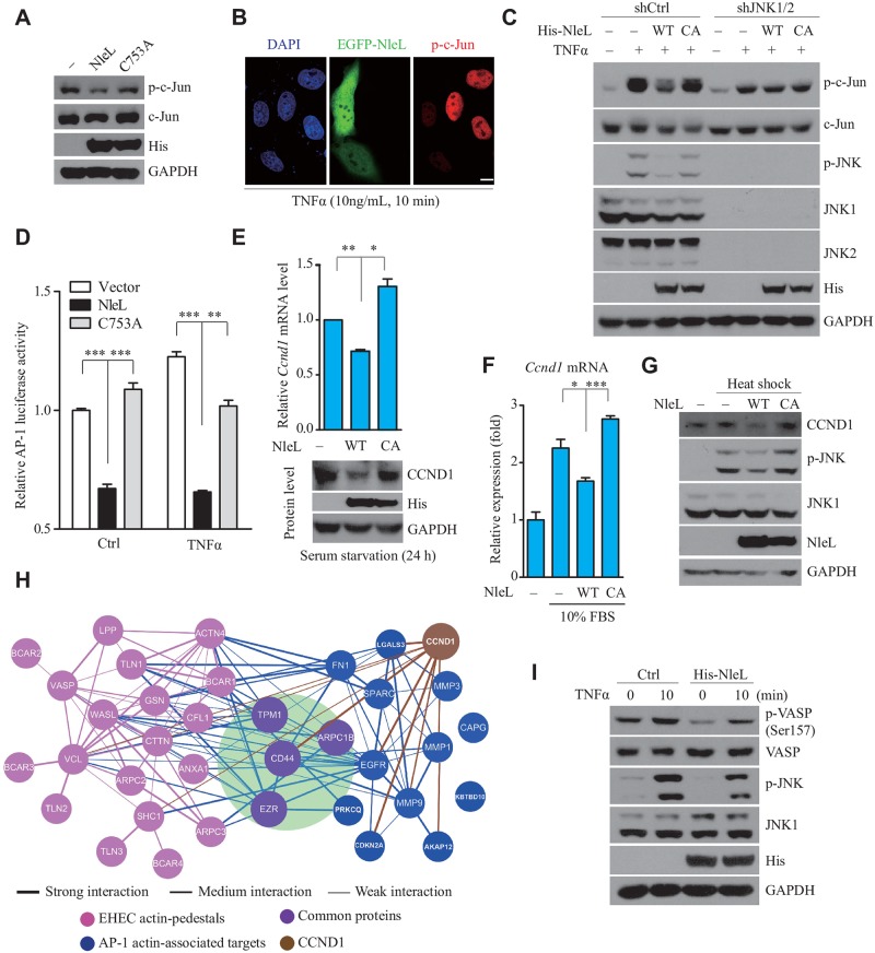
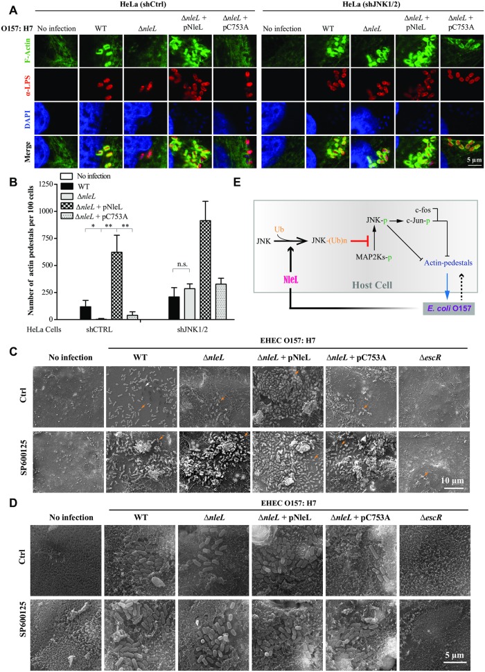
As a major diarrheagenic human pathogen, enterohemorrhagic Escherichia coli (EHEC) produce attaching and effacing (A/E) lesions, characterized by the formation of actin pedestals, on mammalian cells. A bacterial T3SS effector NleL from EHEC O157:H7 was recently shown to be a HECT-like E3 ligase in vitro, but its biological functions and host targets remain elusive. Here, we report that NleL is required to effectively promote EHEC-induced A/E lesions and bacterial infection. Furthermore, human c-Jun NH2-terminal kinases (JNKs) were identified as primary substrates of NleL. NleL-induced JNK ubiquitylation, particularly mono-ubiquitylation at the Lys 68 residue of JNK, impairs JNK's interaction with an upstream kinase MKK7, thus disrupting JNK phosphorylation and activation. This subsequently suppresses the transcriptional activity of activator protein-1 (AP-1), which modulates the formation of the EHEC-induced actin pedestals. Moreover, JNK knockdown or inhibition in host cells complements NleL deficiency in EHEC infection. Thus, we demonstrate that the effector protein NleL enhances the ability of EHEC to infect host cells by targeting host JNK, and elucidate an inhibitory role of ubiquitylation in regulating JNK phosphorylation.


Conflict of interest statement

The authors have declared that no competing interests exist.

Figures

Fig 1
Fig 1. NleL contributes to EHEC attachment to host, and interacts with human JNK1 protein.
(A) Domain structure of NleL from E. coli O157:H7. Cys753 in the C-terminal domain of NleL is the catalytic site responsible for the E3 Ub ligase activity of NleL. (B) Quantification of EHEC O157 attached to HeLa cells by colony formation assay. HeLa cells were infected with indicated EHEC strains with multiplicity of infection (MOI) of 100:1 for 2.5 h, washed with PBS and then re-cultured 2 h in fresh DMEM medium. Infected HeLa cells were thoroughly washed with PBS and aseptically lysed in 1% Triton X-100 buffer. Then bacterial colonies were determined after HeLa lysate being plated onto LB agar. Bars represent mean ± s.d. from three biological replicates, ***P < 0.001 (Student’s t-test, n = 3). (C) NleL promoted attachment of EHEC to mammalian cells. Infected HEK293T cells were thoroughly washed with PBS and subjected to immunofluorescence microscopy with anti-LPS antibody (in red). Scale bar, 50 μm. (D) Interaction of GST-NleL with endogenous JNK1. GST–tagged wild type or C753A mutant of NleL (GST-NleL-CA) were mixed with HEK293T cell lysates and the bound proteins were immunoblotted with the antibody specific for JNK1. GST, GST-tagged wild-type NleL or its C753A mutant were expressed and purified from bacteria. The C753A mutant was enzymatically dead and generated by substitution of the active site Cys753 with Ala. (E) NleL interacted with JNK1 in vivo. Flag-tagged JNK1, His6-tagged NleL (His-NleL) or its C753A mutant (His-NleL-CA) were ectopically expressed in HEK293T cells. Cells were lysed and subjected to immunoprecipitation (IP) with anti-Flag beads, followed by immunoblotting (IB) analysis with indicated antibodies. (F) Human JNK1 interacted with NleL in vitro. His6-tagged JNK1 (His-JNK1), GST-tagged wild-type NleL or its C753A mutant were purified from bacteria. (G) GST pull-down assay of full-length NleL or its truncations with JNK1. GST-tagged full-length NleL or its truncation mutants were individually mixed with lysates of the HEK293T cells expressing JNK1. After pull-down, Flag-tagged JNK1 was detected by IB analysis with anti-Flag. (H) Flag-tagged NleL secreted by EHEC O157 interacted with JNK in HEK293T cells. HEK293T cells were infected by EHEC strain expressing Flag-tagged NleL. Infected cells were thoroughly washed with PBS and lysed in IP buffer, and then IP with anti-JNK antibody and IB with anti-Flag antibody were successively performed. Data shown here are representative of at least three independent experiments.
Fig 2
Fig 2. NleL ubiquitylates human JNK1.
(A) NleL ubiquitylated endogenous JNK during EHEC O157:H7 infection. HEK293T cells were infected with EHEC strains with MOI of 100:1. 2.5 h after infection, cells were washed with PBS and cultured in the fresh DMEM medium for 2 h. Cells were lysed and subjected to IP with anti-JNK antibody and IB with anti-Ub antibody. (B) NleL ubiquitylated JNK1 in vivo. HEK293T cells were co-transfected with plasmids encoding HA-tagged Ub, Flag-tagged JNK1, His6-tagged wild-type NleL or its C753A mutant. Flag-tagged JNK1 was immunoprecipitated with anti-Flag M2 beads in denaturing RIPA buffer, followed by IB analyses with anti-HA antibody. (C) NleL induced mono- and poly-ubiquitylation of JNK1 in vivo. HEK293T cells were co-transfected with plasmids encoding HA-tagged Ub, Flag-tagged JNK1 and His6-tagged NleL. Flag-tagged JNK1 was immunoprecipitated with anti-Flag beads in denaturing RIPA buffer, followed by immunoblotting with anti-Flag antibody. (D) NleL ubiquitylated JNK1 in vitro. Flag-tagged JNK1 was expressed in HEK293T cells and purified with anti-Flag M2 affinity beads, and were subjected to in vitro ubiquitylation reaction mixture with indicated components. After 60 min incubation, the reactions were stopped and subjected to IB analysis with anti-Flag antibody. (E) NleL promoted mono- and poly-ubiquitylation of JNK1 with K29-Ub. HEK293T cells were co-transfected with plasmids encoding HA-tagged Lys 29 only Ub mutant (HA-K29-Ub), Flag-tagged JNK1, His6-tagged wild-type NleL or its C753A mutant. After immunoprecipitation, immunoblotting analyses with indicated antibodies were performed. Usp2cc, the catalytical core of human deubiquitylating enzyme USP2. (F) Mapping the sites for NleL-mediated ubiquitylation of JNK1 through mass spectrometry (MS) analyses. The immunoprecipitated JNK1 from HEK293T cells expressing NleL was subjected to MS analysis to determine the potential sites for NleL-mediated ubiquitylation. (G) A schematic view of the potential ubiquitylation sites of JNK1. The residues K68, K153, K222, and K265 were highlighted in red color to indicate the major ubiquitylation sites. (H) NleL promoted poly-Ub chains on JNK1 at multiple sites. HEK293T cells were transfected with His6-tagged NleL and Flag-tagged wild-type JNK1 or the mutants with Lys-to-Arg substitution at indicated sites. (I) NleL-mediated poly-ubiquitylation of JNK1 was almost abolished by simultaneous Lys-to-Arg substitutions at three major ubiquitylation sites (K153R/ K222R/K265R, 3KR). (J) Mapping the major site(s) for NleL-induced mono-ubiquitylation on JNK1. Flag-tagged wild-type JNK1 or its mutants bearing K-to-R substitution at each potential ubiquitylation site was individually transfected to HEK293T cells with His-NleL and HA-Ub-K29. (K) Lys-to-Arg substitution at Lys 68 (K68R) of JNK1 abolished NleL-induced mono-ubiquitylation of JNK1 in mammalian cells. (L) K68R of JNK1 abolished NleL-mediated JNK1 mono-ubiquitylation in vitro. Flag-tagged wild-type JNK1 or the indicated mutants (K68R and 3KR) were separately expressed in HEK293T cells and purified with anti-Flag M2 affinity beads. Purified JNK1 protein or its mutants was subjected to in vitro ubiquitylation reaction mixture at 37°C for 1h, followed by immunoblotting with indicated antibodies. All blots were representative of at least three independent experiments.
Fig 3
Fig 3. NleL-mediated JNK1 ubiquitylation inhibits phosphorylation of JNK1.
(A) The ΔnleL strain of EHEC O157:H7 induced higher level of JNK phosphorylation than wild-type strain. Cells were infected with indicated EHEC strains with MOI of 100:1 for 2.5 h, and further sub-cultured 2 h in fresh DMEM medium. Then infected cells were lysed and subjected to IB analysis with indicated antibodies (left). The phosphorylated-JNK/ total JNK ratio (right) were calculated by quantifying protein bands. The lane numbers correspond to those shown in the left diagram. Bars represent means ± s.d., *P < 0.05, **P < 0.01, ***P < 0.001 (Student’s t-test, data are from three independent experiments, (n = 3)). (B) Cells were infected by indicated EHEC strains and then subjected to TNFα (10 ng/ml) treatment at the indicated time points. (C and D) NleL inhibited TNFα-mediated JNKs phosphorylation. HEK293T cells were transfected with plasmids encoding His-NleL or C753A mutant for 24 h. Cells were then stimulated by TNFα (1 ng/ml, C or 10 ng/ml, D). (E) Wild-type NleL, but not the C753A mutant, inhibited basal phosphorylation of ectopically expressed JNK1 in HEK293T cells. (F) NleL-induced JNK1 ubiquitylation conversely correlates with JNK1 phosphorylation. Indicated plasmids were transfected to HEK293T cells for 24 h. After transfection, cells were stimulated by TNFα (10 ng/ml) for 10 min and lysed in denatured RIPA buffer. Cell lysate was subjected to anti-Flag IP to enrich JNK1 protein. Phosphorylation and ubiquitylation status of JNK1 was assayed with indicated antibodies. (G) The E3 activity of NleL inhibits JNK activity. Plasmids expressing Flag–tagged JNK1 and His6-tagged NleL or its C753A mutant were co-transfected to HEK293T cells. 24 h after transfection, cells were treated with or without TNFα (10 ng/ml) for 10 min. Cells were then washed once in PBS and lysed in M2 buffer. Cell lysates were subjected to IP with anti-Flag M2 beads overnight. The anti-Flag M2 beads were then washed five times with M2 buffer, twice with 20mM Hepes (pH 7.4) and then subjected to the in vitro kinase assay, using GST-tagged c-Jun (1-79aa) as the substrate. The phosphorylation status of c-Jun was detected by anti-p-c-Jun antibody. (H) NleL did not suppress the TNFα-stimulated phosphorylation of K68R mutant of JNK1. HEK293T cells transiently expressed wild-type Flag-tagged JNK1 or its indicated mutants (K68R and 3KR). Cells were treated with TNFα (10 ng/ml) for 10 min. Phosphorylation of JNK1 was detected by immunoblotting analysis with anti-p-JNK antibody, and the phosphorylated-JNK1/ total JNK1 ratio (right) were further quantitated. The lane numbers correspond to those shown in the left panel. Bars represent means ± s.d., **P < 0.01, n.s., not significant. (Student’s t-test, data are from at least three independent experiments, (n = 3)). (I) NleL had no effect on TNFα-induced phosphorylation of p38 and Erk in mammalian cells. Cells were transfected with or without the plasmids encoding His6-tagged NleL for 24 h, and then subjected to TNFα treatment (10 ng/ml) at indicated times. Phosphorylation status of endogenous p38 or Erk was determined by IB with respective antibodies. Blots are representative of at least three independent experiments.
Fig 4
Fig 4. NleL targets other human JNK family proteins (JNK2 and JNK3).
(A) A schematic view of the sequence homology between human JNK family proteins. The level of sequence homology between JNK1 and JNK2 or JNK3 (90% and 96%, respectively) is indicated. (B) NleL interacted with JNK2. Flag-tagged JNK2 and His6-tagged NleL (His-NleL) or C753A mutant (His-NleL-CA) were ectopically expressed in cells. Cells were lysed and subjected to Co-IP with anti-Flag beads, followed by IB analysis with indicated antibodies. (C) NleL interacted with endogenous JNKs or ectopically expressed JNK3. GST-tagged NleL, its C753A mutant or GST only was individually subjected to the GST pull-down assay with cell lysates of HEK293T cells transfected with (right) or without (left) Flag-tagged JNK3. The bound proteins were immunoblotted with anti-JNK antibody. (D) NleL promotes ubiquitylation of JNK2 (left) or JNK3 (right) in vivo. (E) NleL blocked TNFα-induced JNK1, JNK2 or JNK3 phosphorylation. HEK293T cells expressing Flag-tagged JNK1, JNK2 or JNK3 and His6-tagged wild-type NleL or its C753A mutant were subjected to TNFα treatment (10 ng/ml) at indicated times. Phosphorylation status of JNKs was determined by IB analyses with anti-p-JNK. (F and G) NleL-associated JNK2/3 ubiquitylation conversely correlates with the phosphorylation of JNK2/3. Phosphorylation and ubiquitylation status of JNK2 (F) or JNK3 (G) was assayed with Flag-JNKs enriched from cells expressing wild-type NleL or the C753A mutant, followed by immunoblotting analyses with indicated antibodies. The in vitro kinase assay was performed with Flag-tagged JNK2 (F). All the blots are representative of at least three independent experiments.
Fig 5
Fig 5. NleL mediated mono-ubiquitylation of JNK1 at Lys 68 inactivates JNK1 through disrupting the JNK1-MKK7 interaction.
(A) NleL had little or no effect on MKK7 phosphorylation. HEK293T cells expressing Flag-tagged MKK7 and His-NleL were subjected to IP with anti-Flag, followed by IB with anti-p-MKK7 antibody. (B) NleL disrupted the interaction between JNK1 and endogenous MKK7. HEK293T cells expressing Flag-tagged JNK1 and His6-tagged NleL or the C753A mutant were lysed 24 h after transfection. Co-immunoprecipitation with anti-Flag M2 beads and immunoblotting with anti-MKK7 antibody were performed (left). The MKK7/JNK1 ratios in immunoprecipitate complexes were further determined by quantifying protein bands (right). (C) Co-immunoprecipitation assays of ectopically expressed Flag-tagged MKK7 and HA-tagged JNK1 (left) or JNK2 (right) in the cells expressing His-NleL or C753A. The immuoprecipitates and whole-cell lysates (input) were subjected to immunoblotting with the indicated antibodies. (D) NleL did not disrupt the interaction between JNK1 and MKK4. (E) K68R of JNK1 almost totally abolished the NleL-mediated disruption of the JNK1-MKK7 interaction. HEK293T cells transiently expressed wild-type Flag-tagged JNK1 or the K68R mutant with or without His6-tagged NleL. Cells were treated with TNFα (10 ng/ml) for 10 min before being lysed. Then immunoprecipitation with anti-Flag M2 beads and subsequent immunoblotting with anti-MKK7 antibody were performed. The MKK7/JNK1 ratio in immunoprecipitates (right) were further determined by quantifying the densities of the concerned protein bands. (F) NleL suppressed the interaction between JNK1 and MKK7 by promoting mono-ubiquitylation on Lys 68 of JNK1. HEK293T cells were transfected with indicated plasmids for 24 h, following by TNFα (10 ng/ml) stimulation for 10 min. (G) Co-expression of GST-tagged NleL and His6-tagged JNK1 in bacteria showed no effect on JNK1 phosphorylation by MKK7 in vitro. His6-tagged JNK1 was purified by using Ni-NTA beads from E. coli BL21 (DE3) strain co-expressing His6-tagged JNK1 and GST-tagged NleL or GST only. HEK293T cells expressing Flag-tagged MKK7 (with or without UV stimulation) were lysed in M2 buffer and subjected to anti-Flag immunoprecipitation. Then the beads bounded with Flag-tagged MKK7 were subjected to in vitro kinase assay with bacterially purified His6-tagged JNK1 as the substrate, followed by IB analysis with anti-p-JNK antibody. In (B), (E), Bars represent means ± s.d. from three biological replicates, *P < 0.05, **P < 0.01, n.s., not significant. (Student’s t-test, data are from three independent experiments, (n = 3)). Blots are representative of at least three independent experiments.
Fig 6
Fig 6. NleL-mediated JNK ubiquitylation disrupts the JNK/AP-1 signaling pathway in the mammalian cells.
(A) NleL inhibited the basal-level phosphorylation of c-Jun in HEK293T cells. (B) NleL prevented the activation of endogenous c-Jun upon TNFα stimulation. Cells were transfected with EGFP-NleL for 24 h before TNFα stimulation (10 ng/ml). Immunofluorescence microscopy analysis was performed with anti-phosphorylated c-Jun antibody (p-c-Jun, in red). Nuclei were counterstained with DAPI (blue). Scale bars represent 10 μm. Shown are representative images from three independent experiments. (C) Jnk1/2 knockdown blocked NleL-mediated inactivation of c-Jun. Cells stably expressing control shRNA (shCtrl) or shRNA for Jnk1/2 (shJNK1/2) were transfected with indicated plasmids for 24 h before TNFα stimulation (10 ng/ml, 10min). Then phosphorylation level of c-Jun and JNK were determined by IB analyses. (D) NleL suppressed the transcription factor activity of AP-1. HEK293T cells expressing wild-type NleL or its C753A mutant were co-transfected with the dual luciferase AP-1 reporter system. Twenty-four hours after transfection, the cells were stimulated by TNFα (10 ng/mL, 6 h) and then subjected to a luciferase activity assay. Data are represented as the mean ± s.d. from three biological replicates, ***P<0.001. (Student’s t-test, data from three biological replicates (n = 3)). (E) NleL down-regulated the homeostatic level of endogenous cyclin D1 (CCND1) at mRNA (upside) or protein (downside) level. Data are represented as the mean ± s.d from three biological replicates, *P < 0.05, **P < 0.01. (Student’s t-test). (F) NleL reduced 10% FBS-stimulated transcription of cyclin D1. HEK293T cells expressing NleL or C753A mutant were serum-starved for 24 h and then stimulated with 10% FBS. Then qRT-PCR was performed to determine cyclin D1 expression at mRNA level. Data are shown as the mean ± s.d. from three biological replicates, *P < 0.05, ***P < 0.001 (Student’s t-test, n = 3). (G) NleL suppressed the CCND1 expression up-regulated by heat shock. Cells expressing NleL or C753A were serum-starved for 24 h, and then subjected to heat shock (42°C, 40 min). (H) Protein-protein interactions among the actin-associated proteins targeted by transcription factor AP-1 and proteins characterized in EHEC actin pedestals (blue and pink, respectively). Hits common in both groups are shown in purple, with the interactions directly related to AP-1 targets shown in blue and the interactions only among actin-pedestal proteins in pink. And, CCND1 and its interaction are brown. Line thickness indicates the interaction level of proteins. (I) NleL regulated phosphorylation of an actin-pedestal protein VASP. HEK293T cells transfected with or without a plasmid expressing His6-tagged NleL were stimulated by TNFα (10 ng/ml, 10 min), and then subjected to IB analyses with indicated antibodies. Blots are representative of at least three independent experiments.
Fig 7
Fig 7. NleL promotes the ability of EHEC O157:H7 to cause A/E lesions on mammalian cells by inhibiting JNK activation.
(A and B) NleL promoted the formation of actin pedestals induced by EHEC O157:H7. HeLa cells stably expressing control shRNA (shCtrl) or shRNA for Jnk1/2 (shJNK1/2) were co-cultured with indicated EHEC strains for 2 h, then washed with PBS and further cultured for 2.5 h in the fresh medium. After infection, cells were subjected to immunofluorescence microscopy analysis. Shown are representative cell images where anti-E. coli LPS staining indicates bacteria (red), DAPI staining marks the nucleus (blue) and F-actin denotes the filamentous actin stained by CytoPainter Phalloidin-iFluor 488 Reagent (green) (A). Shown in (B) are numbers of actin pedestals per 100 cells. More than 100 infected cells were examined for each infection experiment and data are represented as mean ± s.d. from three independent experiments. Statistical significance was determined by Student’s t-test. *P < 0.05, **P < 0.01, n.s., not significant. (C) NleL enhances the ability of EHEC O157:H7 to infect Caco-2 monolayer (grown for 6 days) by targeting host JNKs. Caco-2 monolayers (grown for 6 days) treated with DMSO or JNK inhibitor SP600125 (10 μM) were infected with EHEC strains for 2.5 h, then washed with PBS and further cultured for 4 h in fresh medium. After infection, cells were subjected to scanning electron microscopy (SEM) analyses. The white arrow represents an actin pedestal; the orange arrows represent bacteria. (D) NleL promotes the ability of EHEC O157:H7 to form A/E lesions on Caco-2 monolayers (grown for 21 days) by inhibiting JNK proteins. Caco-2 monolayers (grown for 21 days) treated with DMSO or JNK inhibitor SP600125 (10 μM) were infected with EHEC strains for 2.5 h, then washed with PBS and further cultured for 4 h in fresh medium. After infection, cells were subjected to SEM analyses. (E) A schematic diagram showing how NleL-mediated JNK ubiquitylation promotes A/E lesion and EHEC colonization through suppressing JNK phosphorylation and impairing MKK7/JNK/AP-1 signaling.

Similar articles

Cited by

References

    1. Bugarel M, Martin A, Fach P, Beutin L. Virulence gene profiling of enterohemorrhagic (EHEC) and enteropathogenic (EPEC) Escherichia coli strains: a basis for molecular risk assessment of typical and atypical EPEC strains. BMC Microbiol. 2011;11(1):1. - PMC - PubMed
    1. Nataro JP, Kaper JB. Diarrheagenic Escherichia coli. Clin Microbiol Rev. 1998;11(1):142–201. - PMC - PubMed
    1. Gallegos KM, Conrady DG, Karve SS, Gunasekera TS, Herr AB, Weiss AA. Shiga toxin binding to glycolipids and glycans. PLoS one. 2012;7(2):e30368 doi: 10.1371/journal.pone.0030368 - DOI - PMC - PubMed
    1. Campellone KG, Leong JM. Tails of two Tirs: actin pedestal formation by enteropathogenic E-coli and enterohemorrhagic E-coli O157:H7. Curr Opin Microbiol. 2003;6(1):82–90. - PubMed
    1. Goosney DL, DeVinney R, Finlay BB. Recruitment of cytoskeletal and signaling proteins to enteropathogenic and enterohemorrhagic Escherichia coli pedestals. Infect Immun. 2001;69(5):3315–22. doi: 10.1128/IAI.69.5.3315-3322.2001 - DOI - PMC - PubMed

MeSH terms