

Neuroanatomy of the spleen: Mapping the relationship between sympathetic neurons and lymphocytes
- PMID: 28753658
- PMCID: PMC5533443
- DOI: 10.1371/journal.pone.0182416
Neuroanatomy of the spleen: Mapping the relationship between sympathetic neurons and lymphocytes
Abstract
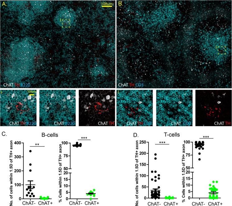
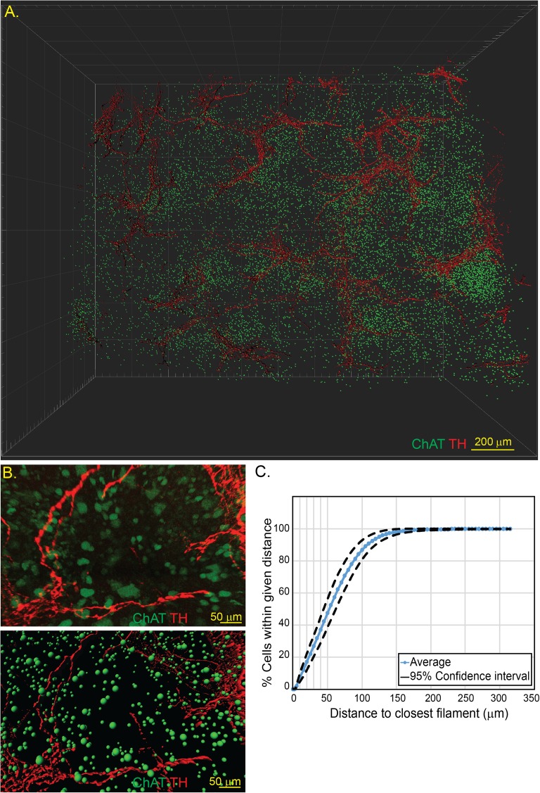
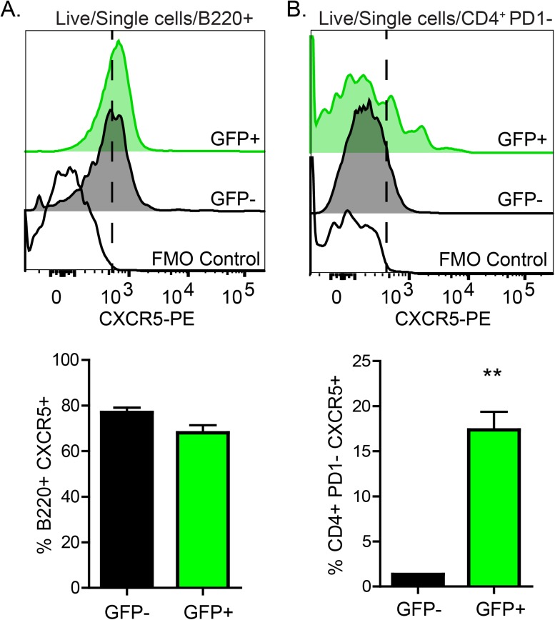
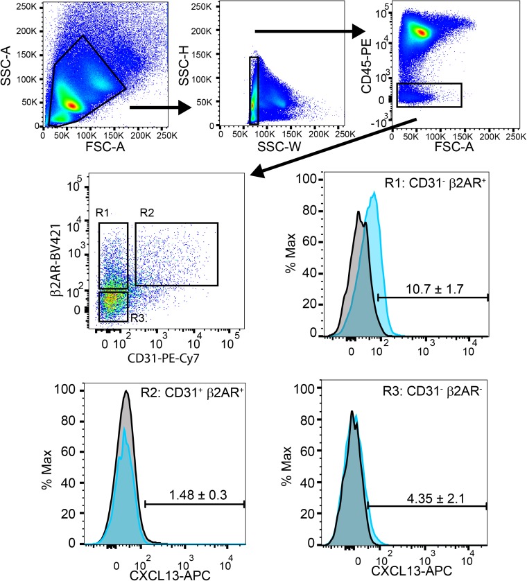
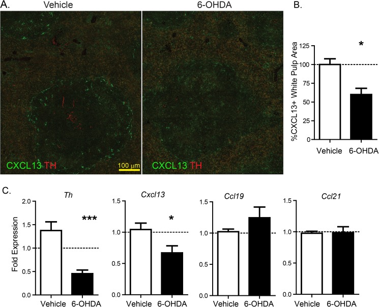
The nervous system plays a profound regulatory role in maintaining appropriate immune responses by signaling to immune cells. These immune cells, including B- and T-cells, can further act as intermediary messengers, with subsets of B- and T-cells expressing choline acetyltransferase (ChAT), the enzyme required for acetylcholine (ACh) synthesis. Neural control of ACh release from ChAT+ T-cells can have powerful immune implications, regulating lymphocyte trafficking, inflammation, and prevent death due to experimental septic shock. Although ACh release from T-cells has been proposed to occur following norepinephrine (NE) released from sympathetic nerve terminals in the spleen, it is unknown how this communication occurs. While it was proposed that tyrosine hydroxylase (TH+) axons form synapse-like structures with ChAT+ T-cells, there is scant evidence to support or refute this phenomenon. With this in mind, we sought to determine the relative abundance of ChAT+ B- and T-cells in close proximity to TH+ axons, and determine what factors contribute to their localization in the spleen. Using confocal microscopy of tissue sections and three-dimensional imaging of intact spleen, we confirmed that ChAT+ B-cells exceed the number of ChAT+ T-cells, and overall few ChAT+ B- or T-cells are located close to TH+ fibers compared to total numbers. The organized location of ChAT+ lymphocytes within the spleen suggested that these cells were recruited by chemokine gradients. We identified ChAT+ B- and T-cells express the chemokine receptor CXCR5; indicating that these cells can respond to CXCL13 produced by stromal cells expressing the β2 adrenergic receptor in the spleen. Our findings suggest that sympathetic innervation contributes to organization of ChAT+ immune cells in the white pulp of the spleen by regulating CXCL13. Supporting this contention, chemical sympathectomy significantly reduced expression of this chemokine. Together, we demonstrated that there does not appear to be a basis for synaptic neuro-immune communication, and that sympathetic innervation can modulate immune function through altering stromal cell chemokine production.
Conflict of interest statement
Figures
References
-
- Muller Paul A, Koscsó B, Rajani Gaurav M, Stevanovic K, Berres M-L, Hashimoto D, et al. Crosstalk between Muscularis Macrophages and Enteric Neurons Regulates Gastrointestinal Motility. Cell. 2014;158(2):300–13. doi: 10.1016/j.cell.2014.04.050 - DOI - PMC - PubMed
-
- Gabanyi I, Muller Paul A, Feighery L, Oliveira Thiago Y, Costa-Pinto Frederico A, Mucida D. Neuro-immune Interactions Drive Tissue Programming in Intestinal Macrophages. Cell. 2016;164(3):378–91. doi: 10.1016/j.cell.2015.12.023 - DOI - PMC - PubMed
-
- Stead RH, Dixon MF, Bramwell NH, Riddell RH, Bienenstock J. Mast cells are closely apposed to nerves in the human gastrointestinal mucosa. Gastroenterology. 1989;97(3):575–85. Epub 1989/09/01. . - PubMed
-
- Riol-Blanco L, Ordovas-Montanes J, Perro M, Naval E, Thiriot A, Alvarez D, et al. Nociceptive sensory neurons drive interleukin-23-mediated psoriasiform skin inflammation. Nature. 2014;510(7503):157–61. doi: 10.1038/nature13199 http://www.nature.com/nature/journal/v510/n7503/abs/nature13199.html#sup.... - DOI - PMC - PubMed
-
- Rosas-Ballina M, Olofsson PS, Ochani M, Valdés-Ferrer SI, Levine YA, Reardon C, et al. Acetylcholine-Synthesizing T Cells Relay Neural Signals in a Vagus Nerve Circuit. Science. 2011. doi: 10.1126/science.1209985 - DOI - PMC - PubMed
MeSH terms
Substances
Grants and funding
LinkOut - more resources
Full Text Sources
Other Literature Sources
