

Quantification of sympathetic hyperinnervation and denervation after myocardial infarction by three-dimensional assessment of the cardiac sympathetic network in cleared transparent murine hearts
- PMID: 28753665
- PMCID: PMC5533449
- DOI: 10.1371/journal.pone.0182072
Quantification of sympathetic hyperinnervation and denervation after myocardial infarction by three-dimensional assessment of the cardiac sympathetic network in cleared transparent murine hearts
Abstract
Background: The sympathetic nervous system is critical in maintaining the normal physiological function of the heart. Its dysfunction in pathological states may exacerbate the substrate for arrhythmias. Obviously, knowledge of its three-dimensional (3D) structure is important, however, it has been revealed by conventional methods only to a limited extent. In this study, a new method of tissue clearance in combination with immunostaining unravels the 3D structure of the sympathetic cardiac network as well as its changes after myocardial infarction.
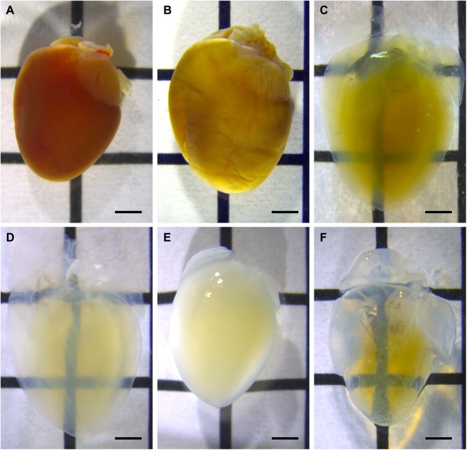
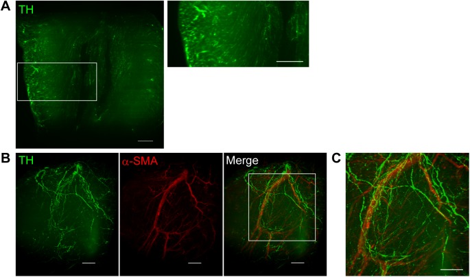
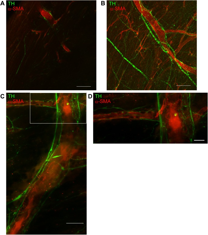
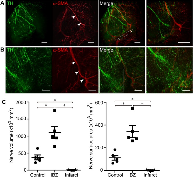
Methods and results: Hearts isolated from adult male mice were optically cleared using the CUBIC-perfusion protocol. After making the hearts transparent, sympathetic nerves and coronary vessels were immunofluorescently labeled, and then images were acquired. The spatial distribution of sympathetic nerves was visualized not only along the epicardial surface, but also transmurally. They were distributed over the epicardial surface and penetrated into the myocardium to twist around coronary vessels, but also independent from the coronary vasculature. At 2 weeks after myocardial infarction, we were able to quantify both denervation distal from the site of infarction and nerve sprouting (hyperinnervation) at the ischemic border zone of the hearts in a 3D manner. The nerve density at the ischemic border zone was more than doubled in hearts with myocardial infarction compared to intact mice hearts (3D analyses; n = 5, p<0.05).
Conclusions: There is both sympathetic hyperinnervation and denervation after myocardial infarction. Both can be visualized and quantified by a new imaging technique in transparent hearts and thereby become a useful tool in elucidating the role of the sympathetic nervous system in arrhythmias associated with myocardial infarction.
Conflict of interest statement
Figures
References
-
- Kawashima T. Anatomy of the cardiac nervous system with clinical and comparative morphological implications. Anatomical science international. 2011;86(1):30–49. doi: 10.1007/s12565-010-0096-0 . - DOI - PubMed
-
- Kaye DM, Lefkovits J, Jennings GL, Bergin P, Broughton A, Esler MD. Adverse consequences of high sympathetic nervous activity in the failing human heart. Journal of the American College of Cardiology. 1995;26(5):1257–63. Epub 1995/11/01. doi: 10.1016/0735-1097(95)00332-0 - DOI - PubMed
-
- Cohn JN, Levine TB, Olivari MT, Garberg V, Lura D, Francis GS, et al. Plasma norepinephrine as a guide to prognosis in patients with chronic congestive heart failure. The New England journal of medicine. 1984;311(13):819–23. Epub 1984/09/27. doi: 10.1056/NEJM198409273111303 . - DOI - PubMed
-
- Jacobson AF, Senior R, Cerqueira MD, Wong ND, Thomas GS, Lopez VA, et al. Myocardial iodine-123 meta-iodobenzylguanidine imaging and cardiac events in heart failure. Results of the prospective ADMIRE-HF (AdreView Myocardial Imaging for Risk Evaluation in Heart Failure) study. Journal of the American College of Cardiology. 2010;55(20):2212–21. doi: 10.1016/j.jacc.2010.01.014 . - DOI - PubMed
-
- Tamaki S, Yamada T, Okuyama Y, Morita T, Sanada S, Tsukamoto Y, et al. Cardiac Iodine-123 Metaiodobenzylguanidine Imaging Predicts Sudden Cardiac Death Independently of Left Ventricular Ejection Fraction in Patients With Chronic Heart Failure and Left Ventricular Systolic Dysfunction. Journal of the American College of Cardiology. 2009;53(5):426–35. doi: 10.1016/j.jacc.2008.10.025 - DOI - PubMed
MeSH terms
LinkOut - more resources
Full Text Sources
Other Literature Sources
Medical
