

GIT1 gene deletion delays chondrocyte differentiation and healing of tibial plateau fracture through suppressing proliferation and apoptosis of chondrocyte
- PMID: 28754105
- PMCID: PMC5534123
- DOI: 10.1186/s12891-017-1653-7
GIT1 gene deletion delays chondrocyte differentiation and healing of tibial plateau fracture through suppressing proliferation and apoptosis of chondrocyte
Abstract
Background: Although tibial plateau fracture is an uncommon injury, its regulation is challenging and there are some influencing factors, including the effects of severe bone displacement, depression and cancellous bone cartilage, and inevitable cartilage damage. And GIT1 plays an important role in bone mass and 78 osteoblast cell migration.
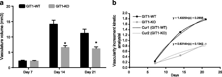
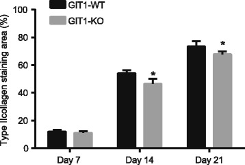
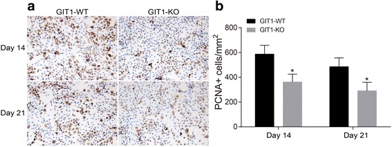
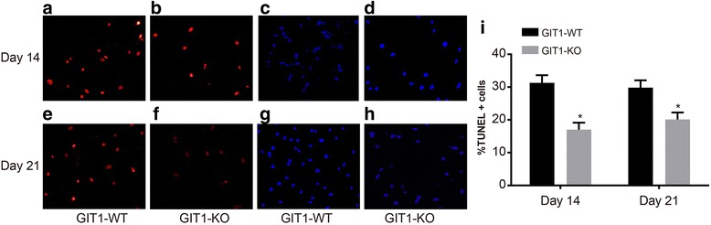
Methods: The study used 72 C57/BL6 mice. A tibial plateau fracture model was established by using mice with the same number of GIT1 gene deletions (the experimental group) and their wild-type littermates (the control group). Joint and bone callus recovery were evaluated by X-ray and CT thin layer scans. Micro CT assay and histomorphometry were conducted in order to evaluate the volume of newly formed blood vessels. Type II collagen expression in tibial tissues after tibial plateau fracture were detected by immunohistochemistry after 7, 14 and 21 days. The number of proliferating cell nuclear antigen (PCNA) positive cells after tibial plateau fracture was tested by immunohistochemistry after 14 and 21 days. The terminal deoxynucleotidyl transferase-mediated dUTP-biotin nick end labeling (TUNEL) staining was conducted after 14 and 21 days in order to test chondrocyte apoptosis in tibial tissues after tibial plateau fracture.
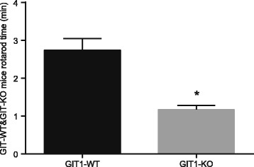
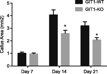
Results: The GIT1 gene deletion group mice spent less time on the rotating rod than the control group mice (P < 0.05). Compared with the control group, postoperative recovery was retarded, because GIT1 gene deletion slowed down neovascularization after tibial plateau fracture (P < 0.05). Compared with the control group, mouse type II collagen expression significantly decreased in the GIT1 gene deletion group, and the proportion of PCNA positive cells significantly decreased (P < 0.05). The TUNEL results indicate that GIT1 gene deletion led to reduced chondrocyte apoptosis.
Conclusion: GIT1 gene deletion can inhibit chondrocyte proliferation and apoptosis during the recovery of tibial plateau fracture, so as to delay chondrocyte differentiation and tibial plateau fracture healing.
Keywords: Apoptosis; GIT1; Gene deletion; Proliferation; Tibial plateau fracture.
Conflict of interest statement
Ethics approval
The study was approved by the Ethics Committee of the Second Hospital of Shandong University. All procedures were in compliance with ethical guidelines for animal experiments. All efforts were made to minimize animal suffering.
Consent for publication
Not applicable.
Competing interests
The authors declare that they have no competing interests.
Publisher’s Note
Springer Nature remains neutral with regard to jurisdictional claims in published maps and institutional affiliations.
Figures
Similar articles
-
Impaired angiogenesis during fracture healing in GPCR kinase 2 interacting protein-1 (GIT1) knock out mice.PLoS One. 2014 Feb 19;9(2):e89127. doi: 10.1371/journal.pone.0089127. eCollection 2014. PLoS One. 2014. PMID: 24586541 Free PMC article.
-
Bax deficiency in mice increases cartilage production during fracture repair through a mechanism involving increased chondrocyte proliferation without changes in apoptosis.Bone. 2008 Nov;43(5):880-8. doi: 10.1016/j.bone.2008.07.239. Epub 2008 Jul 29. Bone. 2008. PMID: 18708175
-
Smoking delays chondrogenesis in a mouse model of closed tibial fracture healing.J Orthop Res. 2006 Dec;24(12):2150-8. doi: 10.1002/jor.20263. J Orthop Res. 2006. PMID: 17013832
-
In situ localization of collagen production by chondrocytes and osteoblasts in fracture callus.J Bone Joint Surg Am. 1989 Jan;71(1):69-77. J Bone Joint Surg Am. 1989. PMID: 2643609 Review.
-
Scaffolds direct Src-specific signaling in response to angiotensin II: new roles for Cas and GIT1.Mol Pharmacol. 2004 Apr;65(4):822-5. doi: 10.1124/mol.65.4.822. Mol Pharmacol. 2004. PMID: 15044610 Review. No abstract available.
Cited by
-
LncRNA HAGLR absorbing miR-214-3p promotes BMP2 expression and improves tibial fractures.Am J Transl Res. 2021 Oct 15;13(10):11065-11080. eCollection 2021. Am J Transl Res. 2021. PMID: 34786043 Free PMC article.
-
Dendronized chitosan hydrogel with GIT1 to accelerate bone defect repair through increasing local neovascular amount.Bone Rep. 2023 Aug 30;19:101712. doi: 10.1016/j.bonr.2023.101712. eCollection 2023 Dec. Bone Rep. 2023. PMID: 37744736 Free PMC article.
-
GIT1 overexpression promotes epithelial-mesenchymal transition and predicts poor prognosis in hepatocellular carcinoma.Bioengineered. 2021 Dec;12(1):30-43. doi: 10.1080/21655979.2020.1855914. Bioengineered. 2021. PMID: 33258389 Free PMC article.
-
Single-cell RNA Seq reveals cellular landscape-specific characteristics and potential etiologies for adolescent idiopathic scoliosis.JOR Spine. 2021 Dec 8;4(4):e1184. doi: 10.1002/jsp2.1184. eCollection 2021 Dec. JOR Spine. 2021. PMID: 35005449 Free PMC article.
-
Inhibiting GIT1 reduces the growth, invasion, and angiogenesis of osteosarcoma.Cancer Manag Res. 2018 Nov 29;10:6445-6455. doi: 10.2147/CMAR.S181066. eCollection 2018. Cancer Manag Res. 2018. PMID: 30555255 Free PMC article.
References
MeSH terms
Substances
LinkOut - more resources
Full Text Sources
Other Literature Sources
Medical
Miscellaneous
