. 2017 Jul 29;36(1):61.
doi: 10.1186/s40880-017-0226-3.

Prognostic value of programmed death-1, programmed death-ligand 1, programmed death-ligand 2 expression, and CD8(+) T cell density in primary tumors and metastatic lymph nodes from patients with stage T1-4N+M0 gastric adenocarcinoma

Affiliations

Prognostic value of programmed death-1, programmed death-ligand 1, programmed death-ligand 2 expression, and CD8(+) T cell density in primary tumors and metastatic lymph nodes from patients with stage T1-4N+M0 gastric adenocarcinoma

Yuan Gao et al. Chin J Cancer. .

Abstract

Background: Anti-programmed death-1/programmed death-ligand 1 (PD-1/PD-L1) immunotherapy has been proved to be effective on gastric cancer in ongoing clinical trials. However, the value of PD-L1 in predicting responses of patients with gastric cancer to anti-PD-1/PD-L1 immunotherapy is controversial. Some studies suggested that intra- and inter-tumoral heterogeneity of PD-L1 expression might explain the controversy. This study aimed to analyze the expression of PD-L1, PD-L2, and PD-1 as well as CD8(+) T-cell density in primary tumors and lymph nodes from patients with stage T1-4N+M0 gastric adenocarcinoma to explore the heterogeneity of PD-1 signaling pathway molecules.

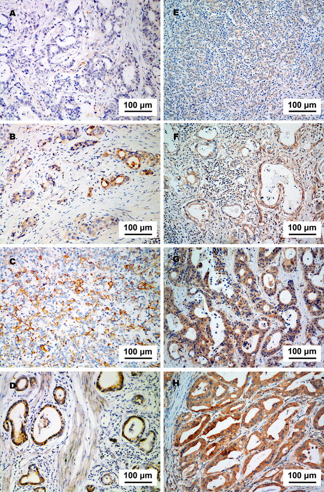
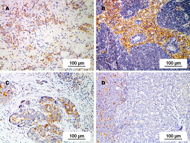
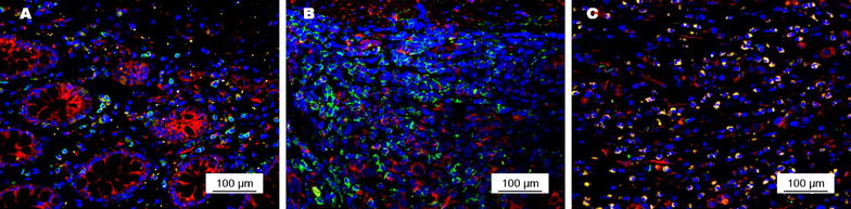
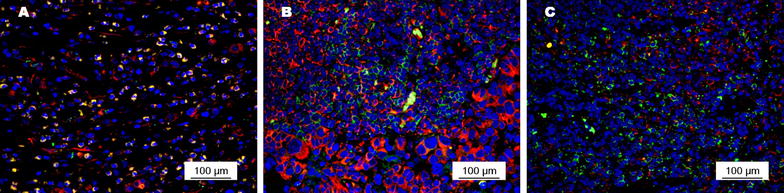
Methods: In primary tumors and metastatic as well as non-metastatic lymph nodes from patients with stage T1-4N+M0 gastric adenocarcinoma, we detected PD-L1 and PD-L2 expression with immunohistochemistry. CD8(+) T-cell density in primary tumors and PD-1 expression on CD8(+) T cells were detected with immunofluorescence. Univariate analysis was used to determine the prognostic values of them. Cox proportional hazard regression model was used to identify independent risk factors that affect patients' overall survival and disease-free survival.

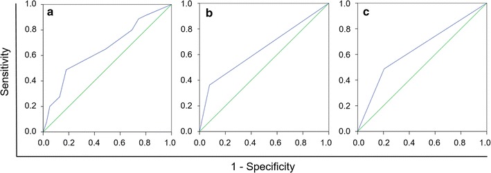
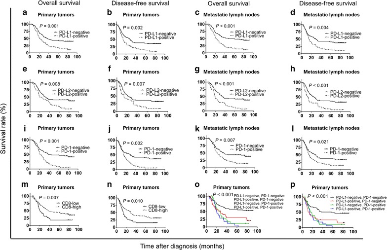
Results: Among 119 eligible patients who had undergone surgical resection, the positive rate of PD-L1 was higher in metastatic lymph nodes than in primary tumors (45.4% vs. 38.7%, P = 0.005); the positive rate of PD-1 on CD8(+) T cells was significantly higher in primary tumors and metastatic lymph nodes than in tumor-free lymph nodes (both P < 0.001). The intensity of PD-1 expression on CD8(+) T cells in primary tumors and in metastatic lymph nodes were stronger than that in tumor-free lymph nodes from the same patient. Beside, the positive rate of PD-L2 did not show any differences between primary tumors and metastatic lymph nodes. In multivariate analysis, PD-L1 expression, PD-L2 expression, a low density of CD8(+) T cells in primary tumors, and PD-1 expression on CD8(+) T cells in primary tumors were associated with poor prognosis.

Conclusion: The expression of PD-L1 is heterogeneous in primary tumors and in metastatic lymph nodes from patients with stage T1-4N+M0 gastric adenocarcinoma, which might explain the inconsistent results in assessing the prognostic value of PD-L1 expression in previous studies.

Keywords: CD8(+) T cells; Gastric cancer; Heterogeneity expression; Prognostic value; Programmed cell death-1; Programmed cell death-ligand 1; Programmed cell death-ligand 2.


Figures

Fig. 1
Fig. 1
Determination of the cut-off scores for programmed cell death-ligand 1 (PD-L1) (a), programmed cell death-ligand 2 (PD-L2) (b), programmed cell death-1 (PD-1) (c) expression with the receiver operating characteristic (ROC) curve analysis
Fig. 2
Fig. 2
PD-L1 and PD-L2 expression in a specimen of gastric adenocarcinoma. a Negative staining of PD-L1. b Weak staining of PD-L1. c Moderate staining of PD-L1. d Strong staining of PD-L1. e Negative staining of PD-L2. f Weak staining of PD-L2. g Moderate staining of PD-L2. h Strong staining of PD-L2
Fig. 3
Fig. 3
PD-L1 expression in primary tumors and corresponding metastatic lymph nodes from individual patients with gastric adenocarcinoma. PD-L1 expression was weaker in the primary tumor (a) than in metastatic lymph nodes (b) of a 71-year-old male patient with stage IIIB gastric adenocarcinoma. PD-L1 expression was stronger in the primary tumor (c) than in metastatic lymph nodes (d) of a 46-year-old female patient with stage IIIB gastric adenocarcinoma
Fig. 4
Fig. 4
PD-1 expression (red signals) on CD8(+) T cells (green signals) in primary tumors of gastric adenocarcinoma. a Moderate intensity of PD-1 expression on CD8(+) T cells with a low density in the primary tumor; b weak intensity of PD-1 expression on CD8(+) T cells with a moderate density in the primary tumor; c strong intensity of PD-1 expression on CD8(+) T cells with a high density in the primary tumor
Fig. 5
Fig. 5
PD-1 expression (red signals) on CD8(+) T cells (green signals) in primary tumors and lymph nodes from a 54-year-old male patient with stage II gastric adenocarcinoma. PD-1 expression on CD8(+) T cells was stronger in the primary tumor (a) and in metastatic lymph nodes (b) than in tumor-free lymph nodes (c)
Fig. 6
Fig. 6
Overall survival (OS) and disease-free survival (DFS) curves of 119 patients with gastric adenocarcinoma according to PD-L1 expression, PD-L2 expression, CD8(+) T-cell density, and PD-1 expression on CD8(+) T cells. a, b OS and DFS curves according to PD-L1 expression status in primary tumors. c, d OS and DFS curves according to PD-L1 expression status in metastatic lymph nodes. e, f OS and DFS curves according to PD-L2 expression status in primary tumors. g, h OS and DFS curves according to PD-L2 expression status in metastatic lymph nodes. i, j OS and DFS curves according to PD-1 expression status on CD8(+) T cells in primary tumors. k, l OS and DFS curves according to PD-1 expression status on CD8(+) T cells in metastatic lymph nodes. m, n OS and DFS curves according to the density of CD8(+) T cells in primary tumors. o, p OS and DFS curves according to both PD-L1 and PD-1 expression in primary tumors

References

    1. Massi D, Brusa D, Merelli B, Falcone C, Xue G, Carobbio A, et al. The status of PD-L1 and tumor-infiltrating immune cells predict resistance and poor prognosis in BRAFi-treated melanoma patients harboring mutant BRAFV600. Ann Oncol. 2015;26(9):1980–1987. doi: 10.1093/annonc/mdv255. - DOI - PubMed
    1. McLaughlin J, Han G, Schalper KA, Carvajal-Hausdorf D, Pelekanou V, Rehman J, et al. Quantitative assessment of the heterogeneity of PD-L1 expression in non-small-cell lung cancer. JAMA Oncol. 2016;2(1):46–54. doi: 10.1001/jamaoncol.2015.3638. - DOI - PMC - PubMed
    1. Ali HR, Glont SE, Blows FM, Provenzano E, Dawson SJ, Liu B, et al. PD-L1 protein expression in breast cancer is rare, enriched in basal-like tumours and associated with infiltrating lymphocytes. Ann Oncol. 2015;26(7):1488–1493. doi: 10.1093/annonc/mdv192. - DOI - PubMed
    1. Choueiri TK, Fay AP, Gray KP, Callea M, Ho TH, Albiges L, et al. PD-L1 expression in nonclear-cell renal cell carcinoma. Ann Oncol. 2014;25(11):2178–2184. doi: 10.1093/annonc/mdu445. - DOI - PMC - PubMed
    1. Massard C, Gordon MS, Sharma S, Rafii S, Wainberg ZA, Luke J, et al. Safety and efficacy of durvalumab (MEDI4736), an anti-programmed cell death ligand-1 immune checkpoint inhibitor, in patients with advanced urothelial bladder cancer. J Clin Oncol. 2016;34(26):3119–3125. doi: 10.1200/JCO.2016.67.9761. - DOI - PMC - PubMed

MeSH terms

Substances