. 2017 Jul 28;14(1):150.
doi: 10.1186/s12974-017-0916-4.

Sensitization of ASIC3 by proteinase-activated receptor 2 signaling contributes to acidosis-induced nociception

Affiliations

Sensitization of ASIC3 by proteinase-activated receptor 2 signaling contributes to acidosis-induced nociception

Jing Wu et al. J Neuroinflammation. .

Abstract

Background: Tissue acidosis and inflammatory mediators play critical roles in pain. Pro-inflammatory agents trypsin and tryptase cleave and activate proteinase-activated receptor 2 (PAR2) expressed on sensory nerves, which is involved in peripheral mechanisms of inflammation and pain. Extracellular acidosis activates acid-sensing ion channel 3 (ASIC3) to trigger pain sensation. Here, we show that a functional interaction of PAR2 and ASIC3 could contribute to acidosis-induced nociception.

Methods: Electrophysiological experiments were performed on both rat DRG neurons and Chinese hamster ovary (CHO) cells expressing ASIC3 and PAR2. Nociceptive behavior was induced by acetic acid in rats.

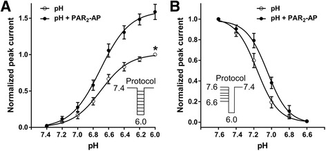
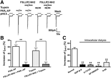
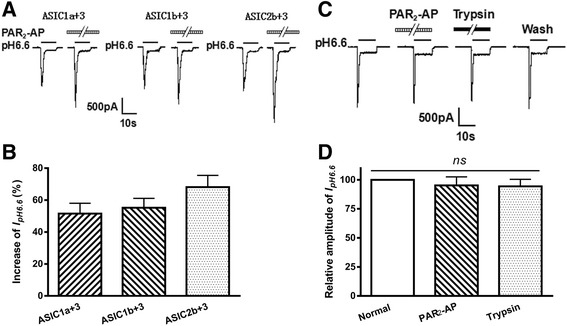
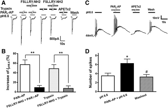
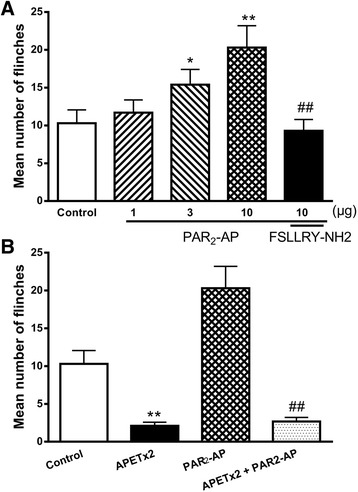
Results: PAR2-AP, PAR2-activating peptide, concentration-dependently increased the ASIC3 currents in CHO cells transfected with ASIC3 and PAR2. The proton concentration-response relationship was not changed, but that the maximal response increased 58.7 ± 3.8% after pretreatment of PAR2-AP. PAR2 mediated the potentiation of ASIC3 currents via an intracellular cascade. PAR2-AP potentiation of ASIC3 currents disappeared after inhibition of intracellular G protein, PLC, PKC, or PKA signaling. Moreover, PAR2 activation increased proton-evoked currents and spikes mediated by ASIC3 in rat dorsal root ganglion neurons. Finally, peripheral administration of PAR2-AP dose-dependently exacerbated acidosis-induced nocifensive behaviors in rats.

Conclusions: These results indicated that PAR2 signaling sensitized ASIC3, which may contribute to acidosis-induced nociception. These represent a novel peripheral mechanism underlying PAR2 involvement in hyperalgesia by sensitizing ASIC3 in primary sensory neurons.

Keywords: Acid-sensing ion channel 3; Dorsal root ganglion neuron; Nociception; Proteinase-activated receptor 2; Proton-gated current.


Conflict of interest statement

Ethics approval and consent to participate

The experimental protocol was approved by the animal research ethics committee of Hubei University of Science and Technology (No. 2016–67).

Consent for publication

Not applicable.

Competing interests

The authors declare that they have no competing interests.

Publisher’s Note

Springer Nature remains neutral with regard to jurisdictional claims in published maps and institutional affiliations.

Figures

Fig. 1
Fig. 1
Potentiation of proton-gated currents by PAR2-AP in CHO cells co-expressing ASIC3 and PAR2. a Representative traces show currents evoked by a pH 6.6 acidic solution for 5 s in CHO cells co-expressing ASIC3 and PAR2. The proton-gated current could be blocked by 500 nM APETx2, an ASIC3 inhibitor. b The sequential current traces illustrate the potentiation of proton-gated currents by different concentrations of PAR2-activating peptide (PAR2-AP: 2-furoyl-LIGRLO-NH2, 10−9–10−4 M). Representative currents were recorded for more than 60 min in a cell with membrane potential clamped at −60 mV. PAR2-AP was pre-applied to external solution for 1 min. c The graph shows PAR2-AP increased the peak amplitude of proton-gated currents in a concentration-dependent manner with an EC50 of 2.9 × 10−7 M. Each point represents the mean ± SEM of 8 to 10 cells
Fig. 2
Fig. 2
Concentration–response relationship for protons and steady-state desensitization of ASIC3 with or without the pre-application of PAR2-AP. a The concentration–response curves for protons with or without 10−5 M PAR2-AP pre-application in CHO cells co-expressing ASIC3 and PAR2. Each point represents the mean ± SEM of 8 to 10 neurons. All current values were normalized to the current response induced by pH 6.0 applied alone (marked with asterisk). The curves shown are a best fit of the data to the logistic equation I = I max/[1 + (pH50/pH)n], where pH is the pH value used, I is the normalized current response value, pH50 is the pH value for half-maximal current response, and n is the Hill coefficient. The curves for protons without and with PAR2-AP pre-application were drawn according to the equation described above. b Steady-state desensitization of homomeric ASIC3 expressed in CHO cells with or without PAR2-AP pre-application. PAR2-AP (10−5 M) induced a rightward shift of the pH dependence of steady-state desensitization. Each point represents the mean ± SEM of 6 to 8 neurons. The holding pH varied from 7.6 to 6.6. All currents were induced by pH 6.0 applied alone
Fig. 3
Fig. 3
The receptor and intracellular mechanisms underlying the potentiation of ASIC3 currents by the activation of PAR2. The a current traces and b bar graphs show that I pH 6.6 was enhanced by PAR2-AP (10−5 M) pre-applied alone for 1 min in CHO cells co-expressing ASIC3 and PAR2. This enhancing effect was inhibited by the co-application of PAR2-AP and FSLLRY-NH2 (10−5 M), a selective PAR2 antagonist. Trypsin, another PAR2 agonist, has a similar increasing effect on I pH 6.6 at concentration of 10−5 M for 1 min. And the enhancing effect of trypsin was also inhibited by FSLLRY-NH2 (10−5 M). Statistical tests were performed using Bonferroni’s post hoc test, and significance is shown as follows: **P < 0.01, compared with white column. n = 10 in each column. The c bar graph shows the percentage increases in the I pH 6.6 induced by PAR2-AP (10−5 M) with recording pipettes filled with the normal internal solution, non-hydrolyzable GDP analog GDP-β-S (500 μM), PLC inhibitor U-73122 (10 μM), PKC inhibitor GF109203X (2 μM), or H-89 (10 μM) containing internal solution. Intracellular dialysis of GDP-β-S, U-73122, GF109203X, or H-89 abolished the enhancing effect of PAR2-AP on I pH 6.6. **P < 0.01, post hoc Bonferroni’s test, compared with normal internal solution. n = 10 in each column
Fig. 4
Fig. 4
PAR2-AP potentiation of proton-gated currents mediated by heteromeric ASIC3 channels. Representative a current traces and b bar graphs show that I pH 6.6 was also enhanced by PAR2-AP (10−5 M) pre-applied for 1 min in CHO cells co-expressing PAR2 and heteromeric ASIC3 plus 1a, 1b, 2a, or 2b channels. n = 8 in each column. The c current traces and d bar graphs show that PAR2-AP and trypsin had no effect on I pH 6.6 in CHO cells expressing alone homomeric ASIC3, but not expressing PAR2. Currents were normalized to control (100%, white column). n = 10 in each column
Fig. 5
Fig. 5
Potentiation of proton-evoked currents and spikes by the activation of PAR2 in rat DRG neurons. The a current traces and b bar graphs show that I pH 6.6 was enhanced by PAR2-AP (10−5 M) or trypsin (10−5 M) pre-applied alone for 1 min in rat DRG neurons. This enhancing effect of PAR2-AP was inhibited by FSLLRY-NH2 (10−5 M), a selective PAR2 antagonist. Also, this proton-induced current could be completely blocked by 2 μM APETx2, an ASIC3 inhibitor. Currents were evoked by extracellular application of a pH 6.6 solution for 5 s in the presence of capsazepine (10 μM) to block proton-induced TRPV1 activation. DRG neurons with membrane potential clamped at −60 mV. The c spike recordings and d bar graphs show that pretreatment of PAR2-AP (10−5 M, for 1 min) increased the acidosis-induced number of action potentials in DRG neurons. The spikes were not evoked by pH 6.6 acidic solution in the presence of 2 μM APETx2. Action potentials were evoked by pH 6.6 acidic solution for 5 s with current clamp recording in the presence of capsazepine (10 μM) to block proton-induced TRPV1 activation. The acidosis-evoked action potentials recovered to control condition after washout of PAR2-AP. *P < 0.05, paired t test, compared with pH 6.6 column alone; #P < 0.05, paired t test, compared with PAR2-AP + pH 6.6 column, n = 9 in each column
Fig. 6
Fig. 6
Effect of PAR2-AP on nociceptive responses to intraplantar injection of acetic acid in rats. The a bar graph shows that the nociceptive responses are evoked by intraplantar injection of acetic acid (30 μl, pH 6.0) in the presence of the TRPV1 inhibitor capsazepine (100 μM). The pretreatment of PAR2-AP increased the flinching behavior induced by acetic acid in a dose-dependent manner (1–10 μg). The effect of PAR2-AP (10 μg) was blocked by co-treatment of FSLLRY-NH2 (20 μg), a selective PAR2 antagonist. *P < 0.05, **P < 0.01, Bonferroni’s post hoc test, compared with control; ##P < 0.01, Bonferroni’s post hoc test, compared with PAR2-AP (10 μg) column. The b bar graph shows that the acidosis-evoked nociception and increased pain response induced by PAR2-AP (10 μg) were blocked by pretreatment with APETx2 (20 μl, 20 μM), an ASIC3 inhibitor. **P < 0.01, Bonferroni’s post hoc test, compared with control; ##P < 0.01, Bonferroni’s post hoc test, compared with PAR2-AP column. Each bar represents the number of flinches that the animals spent licking/lifting the injected paw during first 5-min observation period (mean ± SEM of 10 rats in each group)

Similar articles

Cited by

References

    1. Hollenberg MD, Compton SJ. International Union of Pharmacology. XXVIII. Proteinase-activated receptors. Pharmacol Rev. 2002;54:203–217. doi: 10.1124/pr.54.2.203. - DOI - PubMed
    1. Adams MN, Ramachandran R, Yau MK, Suen JY, Fairlie DP, Hollenberg MD, Hooper JD. Structure, function and pathophysiology of protease activated receptors. Pharmacol Ther. 2011;130:248–282. doi: 10.1016/j.pharmthera.2011.01.003. - DOI - PubMed
    1. Gieseler F, Ungefroren H, Settmacher U, Hollenberg MD, Kaufmann R. Proteinase-activated receptors (PARs)—focus on receptor-receptor-interactions and their physiological and pathophysiological impact. Cell Commun Signal. 2013;11:86. doi: 10.1186/1478-811X-11-86. - DOI - PMC - PubMed
    1. Hollenberg MD, Renaux B, Hyun E, Houle S, Vergnolle N, Saifeddine M, Ramachandran R. Derivatized 2-furoyl-LIGRLO-amide, a versatile and selective probe for proteinase-activated receptor 2: binding and visualization. J Pharmacol Exp Ther. 2008;326:453–462. doi: 10.1124/jpet.108.136432. - DOI - PubMed
    1. Bunnett NW. Protease-activated receptors: how proteases signal to cells to cause inflammation and pain. Semin Thromb Hemost. 2006;32(Suppl 1):39–48. doi: 10.1055/s-2006-939553. - DOI - PubMed

MeSH terms