. 2017 Sep 15:630:38-46.
doi: 10.1016/j.abb.2017.07.015. Epub 2017 Jul 25.

Characterization of secondary structure and lipid binding behavior of N-terminal saposin like subdomain of human Wnt3a

Affiliations

Characterization of secondary structure and lipid binding behavior of N-terminal saposin like subdomain of human Wnt3a

Aparna Krishnamoorthy et al. Arch Biochem Biophys. .

Abstract

Wnt signaling is essential for embryonic development and adult homeostasis in multicellular organisms. A conserved feature among Wnt family proteins is the presence of two structural domains. Within the N-terminal (NT) domain there exists a motif that is superimposable upon saposin-like protein (SAPLIP) family members. SAPLIPs are found in plants, microbes and animals and possess lipid surface seeking activity. To investigate the function of the Wnt3a saposin-like subdomain (SLD), recombinant SLD was studied in isolation. Bacterial expression of this Wnt fragment was achieved only when the core SLD included 82 NT residues of Wnt3a (NT-SLD). Unlike SAPLIPs, NT-SLD required the presence of detergent to achieve solubility at neutral pH. Deletion of two hairpin loop extensions present in NT-SLD, but not other SAPLIPs, had no effect on the solubility properties of NT-SLD. Far UV circular dichroism spectroscopy of NT-SLD yielded 50-60% α-helix secondary structure. Limited proteolysis of isolated NT-SLD in buffer and detergent micelles showed no differences in cleavage kinetics. Unlike prototypical saposins, NT-SLD exhibited weak membrane-binding affinity and lacked cell lytic activity. In cell-based canonical Wnt signaling assays, NT-SLD was unable to induce stabilization of β-catenin or modulate the extent of β-catenin stabilization induced by full-length Wnt3a. Taken together, the results indicate neighboring structural elements within full-length Wnt3a affect SLD conformational stability. Moreover, SLD function(s) in Wnt proteins appear to have evolved away from those commonly attributed to SAPLIP family members.

Keywords: Canonical Wnt signal transduction; Circular dichroism spectroscopy; Limited proteolysis; Liposomes; Saposin; Wnt3a.


Figures

Figure 1
Figure 1. Structural models of Xenopus Wnt8 and NT-SLD
Panel A) Structure of Xenopus Wnt8 complexed with Fzd8-CRD (Protein Data Bank entry: 4FOA) was generated using Visual Molecular Dynamics [47]. The first NT 82 amino acids are in magenta, the SLD is multicolour and the CT domain is in grey. Fzd8-CRD is shown as grey space-fill. The position of 4 tryptophan residues are marked with stars, the remaining tryptophans in the SLD portion of Wnt8 reside in a region masked by the presence of Fzd8-CRD. Panel B) Schematic representation of secondary structure elements of human Wnt3a NT-SLD (adapted from [43]). Cylinders depict α-helices; block arrows indicate β-sheet and dotted lines connect cysteine disulfide bonds. Individual α-helices are color-coded: magenta (N terminal 82 amino acids), cyan (SLD helix a), green (helix b), yellow (helix c) and red (helix d). ‘W’ followed by their sequence position indicates the location of SLD tryptophan residues.
Figure 2
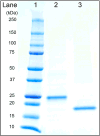
Figure 2. SDS-PAGE of isolated SLD proteins
Lane assignments: lane 1) prestained Precision Plus protein standard, lane 2) NT-SLD (expected molecular weight = 25 kDa) and lane 3) NT-SLD(nh) (expected molecular weight = 20 kDa). Five μg protein was applied to each well and, following electrophoresis, the gel was stained with Gel Code Blue.
Figure 3
Figure 3. Far UV CD spectroscopy of SLDs
Panel A) Spectra of NT-SLD (0.2 mg/ml) in 50 mM Na citrate pH 3, 20 mM NaCl (solid line), 50 mM Na citrate, pH 3, 20 mM NaCl plus 1% SDS (dotted line) or 50 mM sodium citrate, pH 3, 20 mM NaCl plus 50% trifluoroethanol (TFE) (dashed line). Panel B) NT-SLD (solid line) and NT-SLD(nh) (dashed line) in 20 mM NaPO4, pH 7.2, 2% DTAC.
Figure 4
Figure 4. SLD-liposome binding interactions
Liposomes were incubated in the presence or absence of NT-SLD or NT-SLD(nh) at a liposome:protein ratio = 20:1 (w/w). After 1 h incubation, the samples were subjected to sucrose gradient centrifugation. Following centrifugation, five fractions (200 μl each) were collected from top to bottom (1 to 5). The liposome content of each fraction was detected by measuring light scattering intensity at 310 nm and presented graphically (Panel A). An aliquot of each fraction was analyzed by SDS-PAGE and proteins visualized by Imperial protein stain (Panel B). Results presented are representative of two independent experiments.
Figure 5
Figure 5. Fluorescence emission spectra of SLDs
Left) Tryptophan fluorescence emission spectra (excitation 280 nm) of (a) NT-SLD (1 mg/ml) in 10 mM Na phosphate, pH 7, 0.6 M arginine HCl, recorded from 300 to 450 nm in the absence (solid black line) and presence (dotted black line) of 2% DTAC. Right) NT-SLD(nh) (1 mg/ml) in 10 mM Na phosphate, pH 7, 0.6 M arginine HCl, recorded from 300 to 450 nm in the absence (solid blue line) and presence of 2% DTAC (dotted blue line). Fluorescence spectra for NT-SLD and NT-SLD(nh) in 2% DTAC were normalized against respective control samples.
Figure 6
Figure 6. Thrombin-mediated proteolysis of NT-SLDs
NT-SLD and NT-SLD(nh) were incubated in the absence and presence of thrombin in 10 mM Na phosphate, pH 7, 0.6 M arginine HCl, labeled as buffer (panel A) and buffer plus 1% CHAPS (panel B) in final volume of 10 μl for 1, 2 and 4 h at 37°C. Control SLD incubations in the absence of thrombin were incubated for 4 h at 37°C. Following incubation, samples were subjected to SDS-PAGE and stained with Imperial protein stain. The results are representative of two experiments.
Figure 7
Figure 7. Effect of SLDs on L cell viability
Cultured murine L cells were incubated in the absence and presence of increasing concentrations of NT-SLD or NT-SLD(nh) for 24 h. Following incubation, cell viability was measured by the MTT assay. SLDs were presented to the cells in 10 mM Na phosphate, pH 7, 0.6 M arginine HCl. Values reported are the mean ± standard error (n=3).
Figure 8
Figure 8. Effect of SLD on canonical Wnt signaling activity
L cells were incubated with indicated concentrations of Wnt3a and (A) NT-SLD or (B) NT-SLD(nh) for 3 h. Following incubation, the cells were lysed and 10 μg lysate protein applied to a 10% SDS-PAGE gel. Separated proteins were transferred to a PVDF membrane and probed with antibodies directed against β-catenin and GAPDH. The results are representative of three independent experiments.

Similar articles

References

    1. Nusse R. Wnt signaling and stem cell control. Cell Res. 2008;18:523–527. doi: 10.1038/cr.2008.47. - DOI - PubMed
    1. Holstein TW. The evolution of the Wnt pathway. Cold Spring Harb Perspect Biol. 2012;4:a007922. doi: 10.1101/cshperspect.a007922. - DOI - PMC - PubMed
    1. Staal FJT, Luis TC, Tiemessen MM. WNT signalling in the immune system: WNT is spreading its wings. Nat Rev Immunol. 2008;8:581–593. doi: 10.1038/nri2360. - DOI - PubMed
    1. Schulte G. Frizzleds and WNT/β-catenin signaling – The black box of ligand–receptor selectivity, complex stoichiometry and activation kinetics. Eur J Pharmacol. 2015;763:191–195. doi: 10.1016/j.ejphar.2015.05.031. - DOI - PubMed
    1. Angers S, Moon RT. Proximal events in Wnt signal transduction. Nat Rev Mol Cell Biol. 2009;10:468. doi: 10.1038/nrm2717. - DOI - PubMed

Publication types

LinkOut - more resources