

Microhomology-mediated end joining: Good, bad and ugly
- PMID: 28754468
- PMCID: PMC6477918
- DOI: 10.1016/j.mrfmmm.2017.07.002
Microhomology-mediated end joining: Good, bad and ugly
Abstract
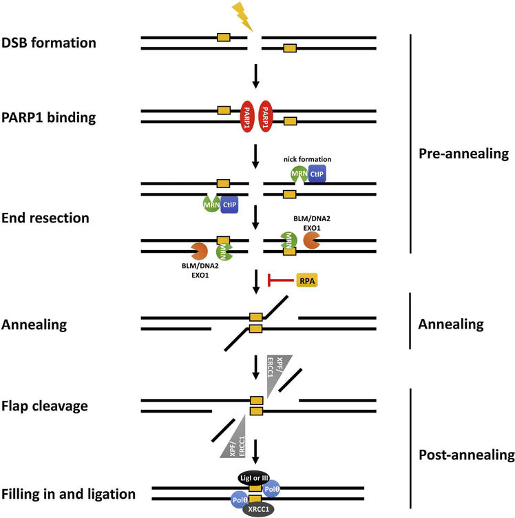
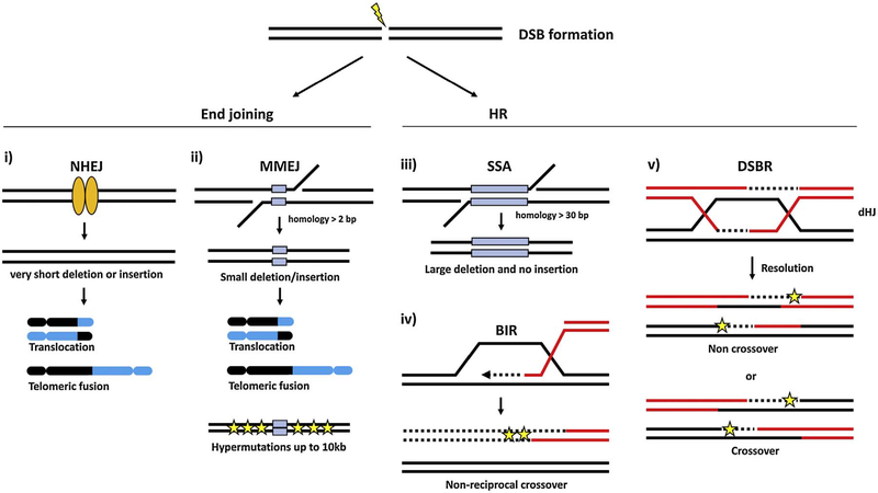
DNA double-strand breaks (DSBs) are induced by a variety of genotoxic agents, including ionizing radiation and chemotherapy drugs for treating cancers. The elimination of DSBs proceeds via distinctive error-free and error-prone pathways. Repair by homologous recombination (HR) is largely error-free and mediated by RAD51/BRCA2 gene products. Classical non-homologous end joining (C-NHEJ) requires the Ku heterodimer and can efficiently rejoin breaks, with occasional loss or gain of DNA information. Recently, evidence has unveiled another DNA end-joining mechanism that is independent of recombination factors and Ku proteins, termed alternative non-homologous end joining (A-NHEJ). While A-NHEJ-mediated repair does not require homology, in a subtype of A-NHEJ, DSB breaks are sealed by microhomology (MH)-mediated base-pairing of DNA single strands, followed by nucleolytic trimming of DNA flaps, DNA gap filling, and DNA ligation, yielding products that are always associated with DNA deletion. This highly error-prone DSB repair pathway is termed microhomology-mediated end joining (MMEJ). Dissecting the mechanisms of MMEJ is of great interest because of its potential to destabilize the genome through gene deletions and chromosomal rearrangements in cells deficient in canonical repair pathways, including HR and C-NHEJ. In addition, evidence now suggests that MMEJ plays a physiological role in normal cells.
Keywords: Chromosome rearrangement; DNA double strand break; End joining; Microhomology; Mutagenesis.
Copyright © 2017 Elsevier B.V. All rights reserved.
Conflict of interest statement
Conflict of interest statement
The authors declare that there are no conflicts of interest.
Figures
Similar articles
-
Rad51 recruitment and exclusion of non-homologous end joining during homologous recombination at a Tus/Ter mammalian replication fork barrier.PLoS Genet. 2018 Jul 19;14(7):e1007486. doi: 10.1371/journal.pgen.1007486. eCollection 2018 Jul. PLoS Genet. 2018. PMID: 30024881 Free PMC article.
-
A role for human homologous recombination factors in suppressing microhomology-mediated end joining.Nucleic Acids Res. 2016 Jul 8;44(12):5743-57. doi: 10.1093/nar/gkw326. Epub 2016 Apr 29. Nucleic Acids Res. 2016. PMID: 27131361 Free PMC article.
-
Ku DNA End-Binding Activity Promotes Repair Fidelity and Influences End-Processing During Nonhomologous End-Joining in Saccharomyces cerevisiae.Genetics. 2018 May;209(1):115-128. doi: 10.1534/genetics.117.300672. Epub 2018 Mar 2. Genetics. 2018. PMID: 29500182 Free PMC article.
-
Mechanisms of DNA double strand break repair and chromosome aberration formation.Cytogenet Genome Res. 2004;104(1-4):14-20. doi: 10.1159/000077461. Cytogenet Genome Res. 2004. PMID: 15162010 Review.
-
Ionizing radiation and genetic risks. XVII. Formation mechanisms underlying naturally occurring DNA deletions in the human genome and their potential relevance for bridging the gap between induced DNA double-strand breaks and deletions in irradiated germ cells.Mutat Res. 2013 Oct-Dec;753(2):114-130. doi: 10.1016/j.mrrev.2013.07.003. Epub 2013 Aug 12. Mutat Res. 2013. PMID: 23948232 Review.
Cited by
-
DNA Damage Response and Immune Defense.Int J Mol Sci. 2020 Oct 12;21(20):7504. doi: 10.3390/ijms21207504. Int J Mol Sci. 2020. PMID: 33053746 Free PMC article. Review.
-
Telomere organization and the interstitial telomeric sites involvement in insects and vertebrates chromosome evolution.Genet Mol Biol. 2022 Nov 14;45(3 Suppl 1):e20220071. doi: 10.1590/1678-4685-GMB-2022-0071. eCollection 2022. Genet Mol Biol. 2022. PMID: 36394537 Free PMC article.
-
Genome-scale CRISPR screens are efficient in non-homologous end-joining deficient cells.Sci Rep. 2019 Oct 31;9(1):15751. doi: 10.1038/s41598-019-52078-9. Sci Rep. 2019. PMID: 31673055 Free PMC article.
-
Harnessing DNA Double-Strand Break Repair for Cancer Treatment.Front Oncol. 2019 Dec 10;9:1388. doi: 10.3389/fonc.2019.01388. eCollection 2019. Front Oncol. 2019. PMID: 31921645 Free PMC article. Review.
-
High-Throughput Analysis of DNA Break-Induced Chromosome Rearrangements by Amplicon Sequencing.Methods Enzymol. 2018;601:111-144. doi: 10.1016/bs.mie.2017.11.028. Epub 2018 Feb 21. Methods Enzymol. 2018. PMID: 29523230 Free PMC article.
References
Publication types
MeSH terms
Substances
Grants and funding
LinkOut - more resources
Full Text Sources
Other Literature Sources
Research Materials
Miscellaneous
