Review
. 2017 Oct;1862(10 Pt B):1221-1232.
doi: 10.1016/j.bbalip.2017.07.009. Epub 2017 Jul 25.

The perilipin family of lipid droplet proteins: Gatekeepers of intracellular lipolysis

Affiliations
Review

The perilipin family of lipid droplet proteins: Gatekeepers of intracellular lipolysis

Carole Sztalryd et al. Biochim Biophys Acta Mol Cell Biol Lipids. 2017 Oct.

Abstract

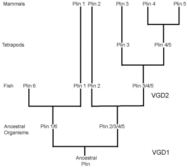
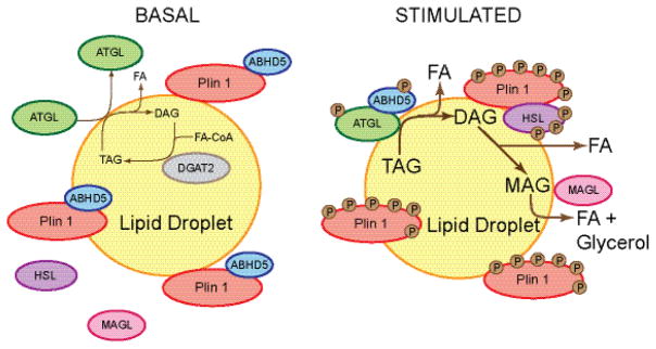
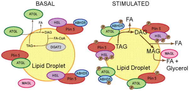
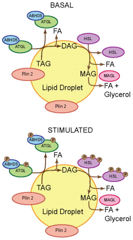
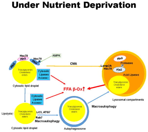
Lipid droplets in chordates are decorated by two or more members of the perilipin family of lipid droplet surface proteins. The perilipins sequester lipids by protecting lipid droplets from lipase action. Their relative expression and protective nature is adapted to the balance of lipid storage and utilization in specific cells. Most cells of the body have tiny lipid droplets with perilipins 2 and 3 at the surfaces, whereas specialized fat-storing cells with larger lipid droplets also express perilipins 1, 4, and/or 5. Perilipins 1, 2, and 5 modulate lipolysis by controlling the access of lipases and co-factors of lipases to substrate lipids stored within lipid droplets. Although perilipin 2 is relatively permissive to lipolysis, perilipins 1 and 5 have distinct control mechanisms that are altered by phosphorylation. Here we evaluate recent progress toward understanding functions of the perilipins with a focus on their role in regulating lipolysis and autophagy. This article is part of a Special Issue entitled: Recent Advances in Lipid Droplet Biology edited by Rosalind Coleman and Matthijs Hesselink.

Keywords: ABHD5; Adipose triglyceride lipase; Autophagy; Hormone-sensitive lipase; Lipid droplet; Lipolysis; Monoacylglycerol lipase; Perilipin; Triacylglycerol.


Conflict of interest statement

Conflict of Interest

The authors, Dawn Brasaemle and Carole Stalryd have no conflicts to disclose

Figures

Figure 1
Figure 1. Theoretical model of perilipin molecular evolution
The perilipins are predicted to have evolved from an ancestral perilipin (Plin) gene expressed in an early chordate. During the first vertebrate genome duplication (VGD) event, two precursor genes that eventually gave rise to perilipins 1 and 6 and perilipins 2, 3, 4, and 5 were formed, followed by a second VGD that gave rise to the individual perilipins 1, 6, 2, and the precursor gene for perilipins 3, 4, and 5. Duplication of the latter precursor gene during the evolution of tetrapods gave rise to perilipin 3 and the precursor gene for perilipins 4 and 5, which diverged during yet another gene duplication event. Perilipin 6 is only expressed in some species of fish, along with perilipins 1, 2, and a precursor gene that gave rise to perilipins 3, 4, and 5 during evolution. Birds and reptiles express perilipins 1, 2, 3, and a perilipin 5-like gene. Only mammals express perilipins 1, 2, 3, 4, and 5. This model was proposed by Granneman, et al. (3), and this figure is derived from Figure 1-figure supplement 2 of that paper. Lines in the model are not to scale and do not represent evolutionary timeframes.
Figure 2
Figure 2. Schematic model of perilipin 1 function in adipocytes under basal and lipolytically stimulated conditions
Under basal conditions, perilipin 1 (Plin 1) resides on lipid droplets in adipocytes and binds ABHD5, thus preventing ABHD5 interaction with and co-activation of ATGL. ATGL, HSL, and presumably MAGL are primarily cytosolic, although low levels of ATGL associate with lipid droplets permitting a low level of basal lipolysis. DAG released from TAG hydrolysis is likely re-esterified by DGAT2 (135). Under lipolytically stimulated conditions, PKA phosphorylates most protein components of the lipolytic machinery, including perilipin 1, ATGL, ABHD5, and HSL, and lipolysis increases markedly. The phosphorylation of perilipin 1 releases phosphorylated ABHD5, which interacts with phosphorylated ATGL to co-activate TAG hydrolysis. Phosphorylated ATGL is highly recruited to lipid droplets where it binds in a perilipin 1-independent manner. Phosphorylated HSL translocates from the cytoplasm to the surfaces of lipid droplets, where it binds to phosphorylated perilipin 1 and hydrolyzes primarily DAG. MAGL cleaves the remaining fatty acid; it is currently unknown whether or not perilipin 1 controls MAGL activity or localization to lipid droplets, or whether the localization of MAGL is altered under stimulated conditions.
Figure 3
Figure 3. Schematic model of perilipin 5 function in myocytes under basal and lipolytically stimulated conditions
Under basal conditions, perilipin 5 (Plin 5) resides primarily on lipid droplets, where it binds HSL, ATGL, and ABHD5, while attenuating lipolysis. Perilipin 5 binding of ATGL and ABHD5 at the carboxyl terminus of perilipin 5 is mutually exclusive; the binding sites overlap, preventing both proteins from binding to a single molecule of perilipin 5, whereas the binding site for HSL is distinct and at the amino terminus. The basal level of TAG hydrolysis catalyzed by ATGL is very low, and released DAG is likely re-esterified by DGAT2. The mechanism for perilipin 5-mediated inhibition of basal lipolysis is unknown, but may include the sequestration of ABHD5, with consequent reduced co-activation of ATGL. Under lipolytically stimulated conditions, PKA phosphorylates most components of the pathway, including perilipin 5, ATGL, CGI-58, and HSL, and lipolysis increases. How these phosphorylation events increase lipolysis is unknown, but may include the release of ABHD5 from the perilipin 5 scaffold to enable its interaction with and co-activation of ATGL. ATGL hydrolyzes TAG; HSL hydrolyzes DAG; MAGL hydrolyzes MAG. It is unknown whether ATGL or HSL remain associated with perilipin 5 under stimulated conditions or how perilipin 5 affects MAGL localization or activity.
Figure 4
Figure 4. Schematic model of perilipin 2 function in cells under basal and lipolytically stimulated conditions
Perilipin 2 localizes almost exclusively to lipid droplets and does not significantly recruit ATGL, ABHD5, or HSL to lipid droplets under either basal or lipolytically stimulated conditions. Perilipin 2 is relatively permissive to lipolysis; lipolysis under basal conditions is approximately comparable to lipolysis when PKA is activated. Perilipin 2 is not a substrate for PKA, although HSL, ATGL, and ABHD5 are substrates. The localization of ATGL with ABHD5 and HSL to perilipin 2 coated lipid droplets is likely transient under both conditions. It is unknown whether or not perilipin 2 affects MAGL localization or activity.
Figure 5
Figure 5. Schematic model of macroautophagy and chaperone-mediated autophagy (CMA) under conditions of nutrient deprivation
Perilipins 2 and 3 provide a barrier to both cytosolic lipolysis and autophagy. Removal of perilipin (Plin) 2 and Plin 3 from the surfaces of lipid droplets is mediated by CMA and precedes lipolysis by both pathways. Plin 2 is phosphorylated by AMP kinase (AMPK) after associating with Hsc70. Plin 2 and Plin 3 are removed as complexes with Hsc70, followed by recognition by Lamp-2A on lysosomes, import into the lumen of the lysosome and degradation. Reduction of Plin 2 and Plin 3 on lipid droplets enables increased binding of cytosolic lipases (such as ATGL), Rab7 and protein effectors of macroautophagy (such as LC3-II) to lipid droplets. This leads to increased release of fatty acids by cytosolic lipases and the engulfment of the lipid droplet by the growing autophagosome. Autophagosomes fuse with lysosomes to form autolysosomes, wherein lysosomal acid lipases hydrolyze triacylglycerols and cholesterol esters, releasing fatty acids and cholesterol. Fatty acids are recycled back into the cytosol to mix with fatty acids generated by cytosolic lipolysis and support mitochondrial β-oxidation.

Similar articles

Cited by

References

    1. Bartholomew SR, Bell EH, Summerfield T, Newman LC, Miller EL, Patterson B, Niday ZP, Ackerman WE, Tansey JT. Distinct cellular pools of perilipin 5 point to roles in lipid trafficking. Biochim Biophys Acta. 2012;1821:268–278. - PMC - PubMed
    1. Hsieh K, Lee YK, Londos C, Raaka BM, Dalen KT, Kimmel AR. Perilipin family members preferentially sequester to either triacylglycerol-specific or cholesteryl-ester-specific intracellular lipid storage droplets. J Cell Sci. 2012;125:4067–4076. - PMC - PubMed
    1. Granneman JG, Kimler VA, Zhang H, Ye X, Luo X, Postlethwait JH, Thummel R. Lipid droplet biology and evolution illuminated by the characterization of a novel perilipin in teleost fish. Elife. 2017;6:e21771. - PMC - PubMed
    1. Blaner WS, O’Byrne SM, Wongsiriroj N, Kluwe J, D’Ambrosio DM, Jiang H, Schwabe RF, Hillman EM, Piantedosi R, Libien J. Hepatic stellate cell lipid droplets: a specialized lipid droplet for retinoid storage. Biochim Biophys Acta. 2009;1791:467–473. - PMC - PubMed
    1. Orban T, Palczewska G, Palczewski K. Retinyl ester storage particles (retinosomes) from the retinal pigmented epithelium resemble lipid droplets in other tissues. J Biol Chem. 2011;286:17248–17258. - PMC - PubMed

Publication types

LinkOut - more resources