

STUB1 regulates TFEB-induced autophagy-lysosome pathway
- PMID: 28754656
- PMCID: PMC5579343
- DOI: 10.15252/embj.201796699
STUB1 regulates TFEB-induced autophagy-lysosome pathway
Abstract
TFEB is a master regulator for transcription of genes involved in autophagy and lysosome biogenesis. Activity of TFEB is inhibited upon its serine phosphorylation by mTOR The overall mechanisms by which TFEB activity in the cell is regulated are not well elucidated. Specifically, the mechanisms of TFEB turnover and how they might influence its activity remain unknown. Here, we show that STUB1, a chaperone-dependent E3 ubiquitin ligase, modulates TFEB activity by preferentially targeting inactive phosphorylated TFEB for degradation by the ubiquitin-proteasome pathway. Phosphorylated TFEB accumulated in STUB1-deficient cells and in tissues of STUB1-deficient mice resulting in reduced TFEB activity. Conversely, cellular overexpression of STUB1 resulted in reduced phosphorylated TFEB and increased TFEB activity. STUB1 preferentially interacted with and ubiqutinated phosphorylated TFEB, targeting it to proteasomal degradation. Consistent with reduced TFEB activity, accumulation of phosphorylated TFEB in STUB1-deficient cells resulted in reduced autophagy and reduced mitochondrial biogenesis. These studies reveal that the ubiquitin-proteasome pathway participates in regulating autophagy and lysosomal functions by regulating the activity of TFEB.
Keywords: TFEB; CHIP (C terminus of HSC70‐Interacting Protein); STUB1; autophagy; lysosomes.
© 2017 The Authors.
Figures
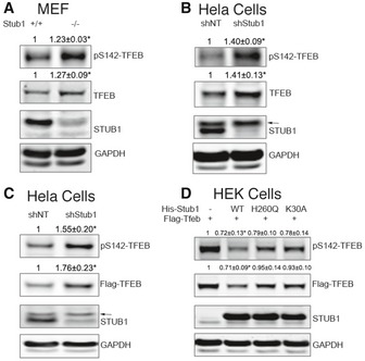
Wild‐type (+/+) or STUB1 knockout (−/−) mouse embryonic fibroblasts (MEFs) were analyzed by Western blot using indicated antibodies.
HeLa cells, stably expressing control shRNA (shNT) or STUB1‐specific shRNA (shStub1), were analyzed by Western blot.
HeLa cells stably expressing shNT or shStub1 were transfected for 48 h with Flag‐TFEB, and cell lysates were analyzed by Western blot.
HEK293 cells were cotransfected for 48 h with Flag‐TFEB together with His‐STUB1, H260Q‐STUB1, or K30A‐STUB1. Cell lysates were then analyzed by Western blot.
- A, B
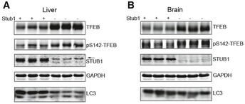
Liver (A) and brain (B) tissues from wild‐type (+/+) or STUB1−/− mice were analyzed by Western blot analysis using indicated antibodies. Arrow denotes a previously described non‐specific band (Sha et al, 2009).
- A, B
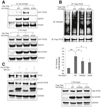
HEK293 cells were cotransfected for 48 h with GFP‐TFEB (A) or Flag‐TFEB (B) together with His‐STUB1, H260Q‐STUB1, or K30A‐STUB1. Cell lysates were then subjected to co‐immunoprecipitation using His (A) or Flag (B) antibodies. The immunocomplexes and input were analyzed by Western blot.
- C
HEK293 cells, cotransfected for 48 h with His‐STUB1 and GFP‐TFEB, were treated for 4 h with 50 μM MG132 or vehicle only. Cell lysates were analyzed by Western blot.
- A
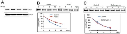
HeLa cells were treated for 2 h with proteasome inhibitor MG132 (50 μM), and cell lysates were analyzed by Western blot.
- B, C
HeLa cells were mock‐treated or treated with proteasome inhibitor PS341 (10 μM; B), or the lysosome inhibitor bafilomycin (10 μM; C), and TFEB half‐life was measured using pulse‐chase analysis. Data are mean ± SD, n = 3. * denotes P < 0.05 using two‐way ANOVA.
- A
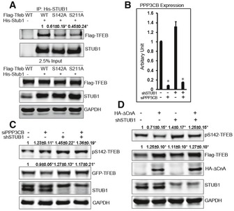
HEK293 cells were cotransfected for 48 h with His‐STUB1 together with Flag‐TFEB, S142A‐TFEB, or S211A‐TFEB. Cell lysates were subjected to co‐immunoprecipitation using His antibodies. The immunocomplexes and input were analyzed by Western blot.
- B, C
HeLa cells stably expressing control shRNA or STUB1‐specific shRNA were transfected for 72 h with non‐target siRNA or PPP3CB siRNA. Cell lysates were then analyzed by real‐time (RT)‐PCR using PPP3CB‐specific primers (B) or by Western blot (C).
- D
Flag‐TFEB expressing HeLa cells were stably transfected with control shRNA or STUB1‐specific shRNA. Cells were transfected for 48 h with vector only or ∆CnA (constitutively active calcineurin), and cell lysates were analyzed by Western blot.
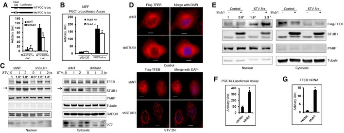
HeLa cells, stably expressing control shRNA (shNT) or STUB1‐specific shRNA (shStub1), were transfected with wild‐type PGC1α promoter–luciferase vector or Clear site mutated PGC1α promoter–luciferase vector and luciferase activity was assayed 24 h post‐transfection.
Wild‐type (+/+) or STUB1−/− MEFs were transfected with vector only or PGC1α promoter–luciferase vector, and luciferase activities were assayed 24 h post‐transfection.
HeLa cells stably expression shNT or shSTUB1 were mock‐treated, starved for 1 or 2 h, and cell lysates were fractionated into nuclear and cytoplasmic fractions and analyzed by Western blot. Arrows denote a previously described non‐specific band (Sha et al, 2009).
HeLa cells stably expressing Flag‐TFEB were transfected for 72 h with shNT or shSTUB1. Cells were mock‐treated or starved for 2 h and analyzed by immunofluorescence using Flag antibodies. Scale bar, 10 μm.
HeLa cells, stably expressing Flag‐TFEB, were transfected with vector only or STUB1. Cells were mock‐treated or starved for 8 or 12 h, and cell lysates (nuclear and cytoplasmic fractions) were analyzed by Western blot.
HeLa cells were transfected for 24 h with vector only or STUB1. Cells were then transfected, for another 24 h, with PGC1α‐luciferase, and luciferase activity assay was performed.
HeLa cells were transfected for 48 h with vector only or STUB1, and TFEB mRNA expression was assayed by RT–PCR.
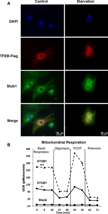
TFEB colocalizes with STUB1 in cytosol. HeLa cells stably expressing Flag‐TFEB were mock‐treated or starved for 2 h and analyzed by immunofluorescence using Flag or Stub1 antibodies. Scale bar, 20 μm.
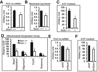
STUB1 deficiency reduced mitochondrial biogenesis. Wild‐type (+/+) or STUB1−/− MEFs were subjected to mitochondrial respiration analysis.
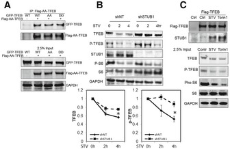
Flag‐AA‐TFEB (TFEB‐S142A/S211A) was cotransfected with GFP‐TFEB, GFP‐AA‐TFEB, or GFP‐DD‐TFEB (TFEB‐S142D/S211D) into HEK293 cells. Cell lysates were subjected to immunoprecipitation using Flag antibodies. Immunocomplexes or 2.5% input were analyzed by Western blot.
HeLa cells stably expressing Flag‐TFEB were transfected for 72 h with shNT or shSTUB1. Cells were mock‐treated or starved for 2 or 4 h and analyzed by Western blot. Graphs show quantitative analysis of TFEB and P‐TFEB.
HeLa cells stably expressing Flag‐TFEB were subjected for 2 h to mock treatment, starvation, or 250 nM Torin1 treatment. Cell lysates were subjected to immunoprecipitation analysis using Flag antibodies.
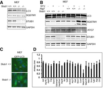
MEFs were analyzed by Western blot.
MEFs were mock‐treated or starved for 2 h (STV) and then analyzed by Western blot. Wild‐type or ATG7−/− MEFs were used as control for autophagy flux analysis.
MEFs were transfected with GFP‐LC3 for 48 h and then treated with chloroquine (50 μm) for 2 h before they were fixed in 4% PFA for fluorescence microscopy. Scale bar, 10 μm.
HeLa cells, stably expressing control shRNA (shNT) or STUB1‐specific shRNA (shStub1), were subjected to RT–PCR‐based gene expression array analysis. Genes that were significantly reduced (P < 0.05 in Student's t‐test analysis) in STUB1 knockdown cells are shown. Data are mean ± SD, n = 3.
- A–D
Wild‐type (+/+) or STUB1−/− MEFs were subjected to RT–PCR analysis of PGC1α (A), mitochondrial copy number analysis (B), ATP content analysis (C), or mitochondrial respiration analysis (D).
- E, F
HeLa cells, stably expressing control shRNA (shNT) or STUB1‐specific shRNA (shStub1), were subjected to RT–PCR analysis of PGC1α (E) or ATP content analysis (F).
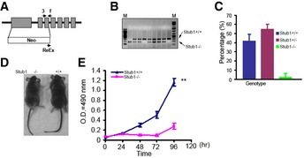
Schematic diagram of generating STUB1 knockout (−/−) mice. Arrows denotes the primers used for genotyping the transgenic mice.
Genotyping of STUB1−/− mice using PCR.
Homozygous STUB1 knockout mice are significantly lower than the expected Mendelian inheritance.
STUB1−/− mice are smaller than wild‐type (+/+) mice.
Proliferation assay of MEFs from STUB1−/− and +/+ mice.
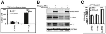
Cells were transfected for 24 h with vector only (control) or TFEB‐S142A/S211A mutant (Flag‐AA‐TFEB) and then transfected for another 24 h with PGC1α promoter–luciferase before luciferase activity was analyzed.
Cells were transfected for 48 h with vector only or Flag‐AA‐TFEB, and cell lysates were analyzed by Western blot.
Cells were treated with transfection reagent only (control) or transfected with vector only or Flag‐AA‐TFEB. ATP content in cell lysates was analyzed 48 h post‐transfection.
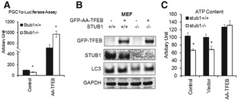
MEFs were transfected for 24 h with vector only (control) or TFEB‐S142A/S211A mutant (GFP‐AA‐TFEB) and then transfected for another 24 h with PGC1α promoter–luciferase before luciferase activity was analyzed.
MEFs were transfected for 48 h with vector only or GFP‐AA‐TFEB, and cell lysates were analyzed by Western blot.
MEFs were treated with transfection reagent only (control) or transfected with vector only or GFP‐AA‐TFEB. ATP content in cell lysates was analyzed 48 h post‐transfection.
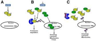
In a nutrient‐rich environment, activated mTOR phosphorylates TFEB, resulting in reduced TFEB activity.
Under conditions of mTOR inhibition, such as during starvation, STUB1 targets phosphorylated TFEB for proteasomal degradation. Non‐phosphorylated TFEB translocates to the nucleus exerting its transcriptional activity promoting genes of the autophagy–lysosomal and mitochondrial pathways as well as TFEB itself.
In STUB1‐deficient cells, phosphorylated TFEB is not efficiently degraded. Accumulating phosphorylated TFEB is inactive and it further reduces TFEB activity by forming heterodimers with non‐phosphorylated TFEB, leading to reduced TFEB translocation to the nucleus. Reduced TFEB activity, in STUB1‐deficient cells, leads to inhibition of autophagy–lysosome pathway and mitochondrial biogenesis.
References
-
- Austin S, St‐Pierre J (2012) PGC1α and mitochondrial metabolism–emerging concepts and relevance in ageing and neurodegenerative disorders. J Cell Sci 125(Pt 21): 4963–4971 - PubMed
-
- Bjørkøy G, Lamark T, Pankiv S, Øvervatn A, Brech A, Johansen T (2009) Monitoring autophagic degradation of p62/SQSTM1. Methods Enzymol 452: 181–197 - PubMed
-
- Dahout‐Gonzalez C, Nury H, Trézéguet V, Lauquin GJ, Pebay‐Peyroula E, Brandolin G (2006) Molecular, functional, and pathological aspects of the mitochondrial ADP/ATP carrier. Physiology 21: 242–249 - PubMed
-
- Fisher DE, Carr CS, Parent LA, Sharp PA (1991) TFEB has DNA‐binding and oligomerization properties of a unique helix‐loop‐helix/leucine‐zipper family. Genes Dev 5: 2342–2352 - PubMed
Publication types
MeSH terms
Substances
Grants and funding
LinkOut - more resources
Full Text Sources
Other Literature Sources
Molecular Biology Databases
Miscellaneous
