. 2017 Sep 1;199(5):1696-1705.
doi: 10.4049/jimmunol.1700551. Epub 2017 Jul 28.

Adaptive from Innate: Human IFN-γ+CD4+ T Cells Can Arise Directly from CXCL8-Producing Recent Thymic Emigrants in Babies and Adults

Affiliations

Adaptive from Innate: Human IFN-γ+CD4+ T Cells Can Arise Directly from CXCL8-Producing Recent Thymic Emigrants in Babies and Adults

Abhishek Das et al. J Immunol. .

Abstract

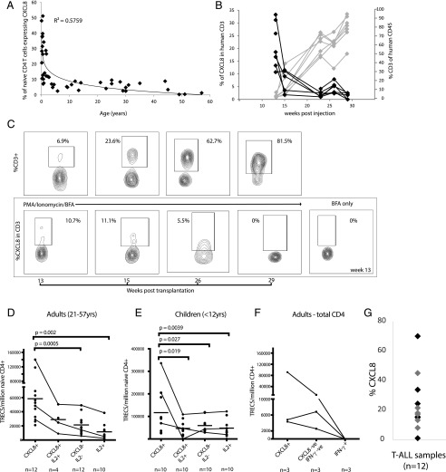
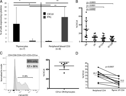
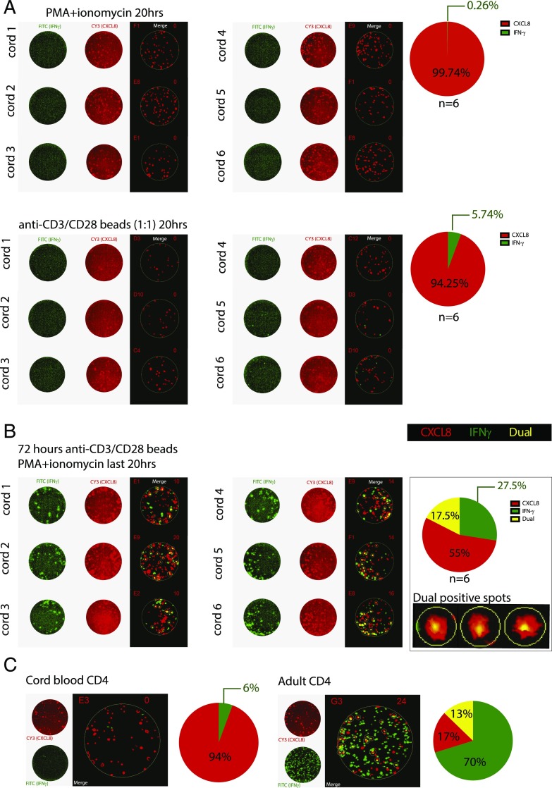
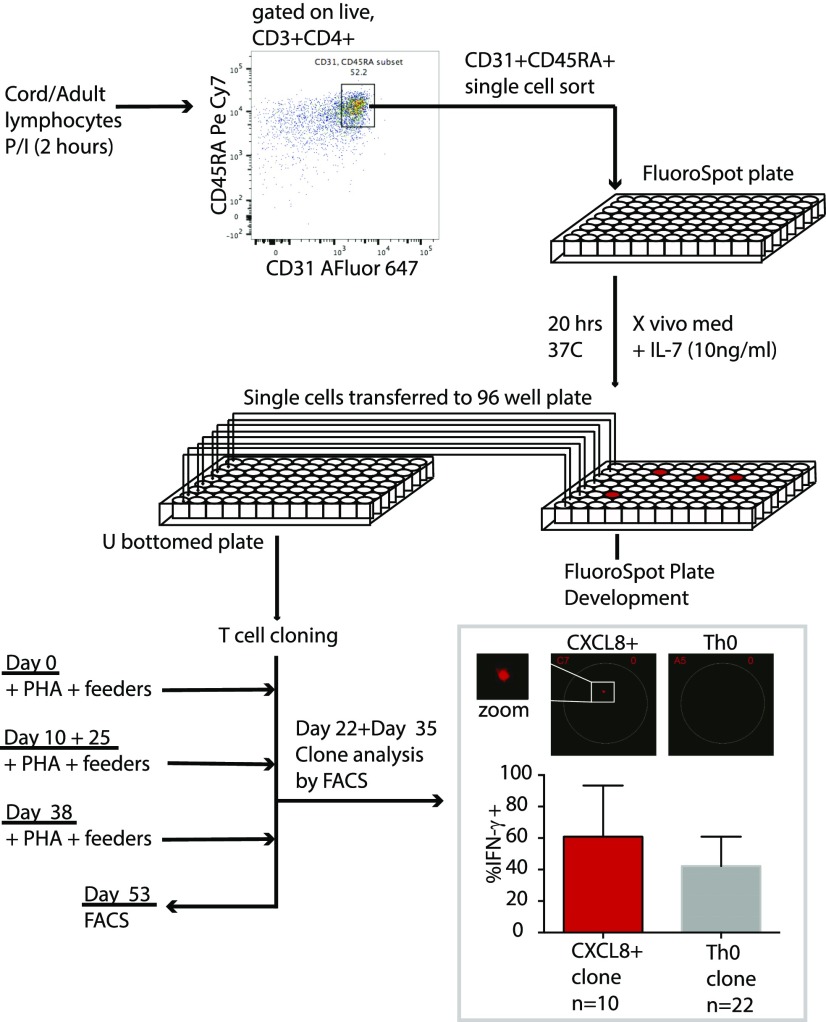
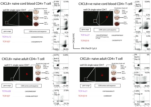
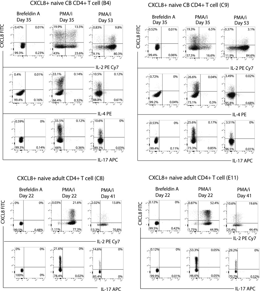
We recently demonstrated that the major effector function of neonatal CD4+ T cells is to produce CXCL8, a prototypic cytokine of innate immune cells. In this article, we show that CXCL8 expression, prior to proliferation, is common in newly arising T cells (so-called "recent thymic emigrants") in adults, as well as in babies. This effector potential is acquired in the human thymus, prior to TCR signaling, but rather than describing end-stage differentiation, such cells, whether isolated from neonates or adults, can further differentiate into IFN-γ-producing CD4+ T cells. Thus, the temporal transition of host defense from innate to adaptive immunity is unexpectedly mirrored at the cellular level by the capacity of human innate-like CXCL8-producing CD4+ T cells to transition directly into Th1 cells.


Figures

FIGURE 1.
FIGURE 1.
CXCL8-producing T cells decline with age in humans and in vivo in humanized mice and are enriched in RTEs. (A) CXCL8 production was determined in naive CD4+ T cells (4 h PI + BFA) obtained from 45 individuals (aged between 3 mo and 57 y). (B) Irradiated NSG mice were reconstituted with human CD34+ cells (CB derived). The graph shows reconstitution of T cells over time in individual mice (gray lines), with a reciprocal decline in T cell production of CXCL8 (black lines). (C) FACS plots show an example of this reciprocal change in one reconstituted NSG mouse; percentage of cells expressing CD3 within human CD45+ cells (upper panels) and percentage of cells expressing CXCL8 among CD3 T cells (lower panels; PI + BFA or BFA alone for 4 h). CD4+ T cells from adults (D and F) or children aged 1–12 y (E) were activated with PI (4 h), and cytokine production was determined by intracellular staining. Individual (cytokine+) subsets were then sorted, and TREC content was determined by qPCR. Results are shown as TREC levels per million naive CD4+ cells or TREC levels per million total CD4+ cells in (F), because very few naive CD4+ cells express IFN-γ. Trend lines depict the differences in TREC levels among the four sorted populations in three patients. (G) CXCL8 production was determined following activation in vitro with PI (4 h, in the presence of BFA) in primary T-ALL samples (n = 12); later stages (T-III/T-IV) are represented by gray diamonds.
FIGURE 2.
FIGURE 2.
CXCL8 production is imprinted in the thymus. CXCL8 production was determined following activation in vitro with PI (4 h, in the presence of BFA) in whole thymocytes versus peripheral blood from humanized mice (37 wk post–hematopoietic stem cell inoculation, n = 7) (A), sorted human thymocyte subsets (n = 15, CD4CD8 DN, CD4+CD8+ double positive, CD4CD8+ SP CD8, CD4+CD8 SP CD4+) (B), and enriched T-lineage–committed DN thymocytes (defined as CD4CD8CD34+CD7+CD5+CD1a+, n = 6) (C). Example and cumulative data are shown. Stimulation: BFA only in dark gray and PI+BFA in light gray. (D) Comparison of CXCL8 production between paired human SP CD4+ thymocytes and naive (CD31+CD45RA+) peripheral CD4+ T cells (n = 10). *p < 0.05, **p < 0.001.
FIGURE 3.
FIGURE 3.
Results of single-cell RNA sequencing. (A) PC analysis of expression data. Each point represents a sample whose expression data has been projected onto the first two PCs for the expression data subset that includes all genes with statistically significant differential expression. (B) Genes providing the largest contribution to weightings for PC1.
FIGURE 4.
FIGURE 4.
CXCL8+ CD4+ T cells are precursors for conventional Th1 effectors. CXCL8/IFN-γ production on a per-cell basis is shown by the FluoroSpot assay in which green spots represent IFN-γ production, and red spots represent CXCL8 production. A merged image to show dual-positive spots is shown for each case; the number of dual-positive spots in the well is annotated on the top right of each image. Corresponding pie charts depict the percentage of IFN-γ+ (green), CXCL8+ (red), and CXCL8+IFN-γ+ (yellow) spots from all of the samples. Naive CD4+ T cells isolated from six cord samples were stimulated ex vivo (20 h PI or anti-CD3+CD28 beads) (A) and for 72 h (anti-CD3+28 beads, followed by PI restimulation for the last 20 h) (B). All experiments were performed in duplicate at a range of cell titrations. All wells depicted had between 7.5 and 22.5 × 10³ cells per well. Examples of dual-positive spots are shown at a higher magnification (right panel). (C) Overnight PI stimulation (10,000 sorted CD4+ T cells per well) for cord and for adult CD4+ T cells. Corresponding pie charts depict the percentage of IFN-γ+ (green), CXCL8+ (red), and CXCL8+IFN-γ+ (dual-yellow) spots.
FIGURE 5.
FIGURE 5.
Schematic diagram demonstrating how single-cell sorting in FluoroSpot plates was performed. Inset: production of IFN-γ is shown for 10 clones derived from CXCL8+ cells and 22 clones derived from Th0 cells, with a representative FluoroSpot well shown above for a CXCL8+ well and a Th0 well. Clones were harvested after sufficient cell numbers had been reached and were stimulated with PI for 4 h prior to staining and acquisition.
FIGURE 6.
FIGURE 6.
Clones derived from a single CXCL8-producing cell produce IFN-γ. Activated (2 h PI) naive CD4+ T cells were single-cell sorted directly into 96-well FluoroSpot plates. Single cells were recovered from the FluoroSpot plate and added to PHA (4 μg/ml) and irradiated feeder cells for cloning. The FluoroSpot plate was developed to reveal a signal left by a single CXCL8-producing cell. Cells cloned were subsequently stimulated (PI; 4 h) at two time points, and dot plots depicting IFN-γ (x-axis) and CXCL8 (y-axis) production at each time point are shown. To show clonality, sequencing of TCRα- and β-chains was performed using DNA extracted from sorted live 7-aminoactinomycin DCD3+CD4+ T cells. The amino acid sequence for the CDR3 region and gene usage are shown for each clone.
FIGURE 7.
FIGURE 7.
Effector function of clones derived from single CXCL8-producing T cells. Naive CD4+ T cells were single cell sorted into FluoroSpot plates for 20 h (2 h preincubation with PI), following by transfer to 96-well plates and cloning by addition of irradiated feeder cells and PHA (4 μg/ml). Once the FluoroSpot plate was developed, it was possible to determine which wells had only a single CXCL8+ spot. Subsequent effector function (IL-4/IL-2/IL-17A) in clones from these CXCL8-producing cells was then tracked over two time points by FACS. Two CB clones and two adult clones are shown.

References

    1. PrabhuDas M., Adkins B., Gans H., King C., Levy O., Ramilo O., Siegrist C. A. 2011. Challenges in infant immunity: implications for responses to infection and vaccines. Nat. Immunol. 12: 189–194. - PubMed
    1. Gibbons D., Fleming P., Virasami A., Michel M. L., Sebire N. J., Costeloe K., Carr R., Klein N., Hayday A. 2014. Interleukin-8 (CXCL8) production is a signatory T cell effector function of human newborn infants. Nat. Med. 20: 1206–1210. - PubMed
    1. Luster A. D. 1998. Chemokines--chemotactic cytokines that mediate inflammation. N. Engl. J. Med. 338: 436–445. - PubMed
    1. Zlotnik A., Yoshie O. 2012. The chemokine superfamily revisited. Immunity 36: 705–716. - PMC - PubMed
    1. Mold J. E., Venkatasubrahmanyam S., Burt T. D., Michaëlsson J., Rivera J. M., Galkina S. A., Weinberg K., Stoddart C. A., McCune J. M. 2010. Fetal and adult hematopoietic stem cells give rise to distinct T cell lineages in humans. Science 330: 1695–1699. - PMC - PubMed

Publication types