. 2017 Sep 1;199(5):1805-1816.
doi: 10.4049/jimmunol.1700515. Epub 2017 Jul 28.

Nitric Oxide Modulates Macrophage Responses to Mycobacterium tuberculosis Infection through Activation of HIF-1α and Repression of NF-κB

Affiliations

Nitric Oxide Modulates Macrophage Responses to Mycobacterium tuberculosis Infection through Activation of HIF-1α and Repression of NF-κB

Jonathan Braverman et al. J Immunol. .

Abstract

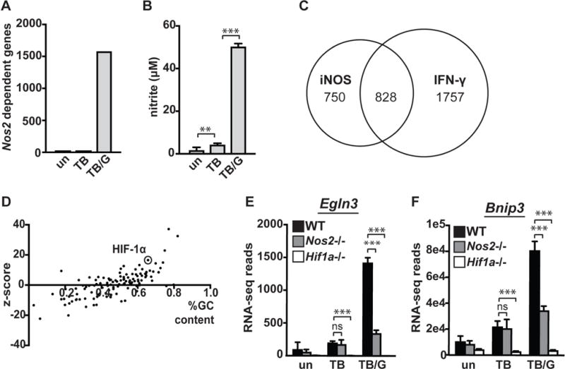
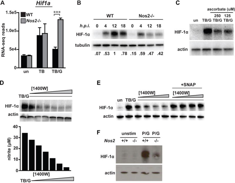
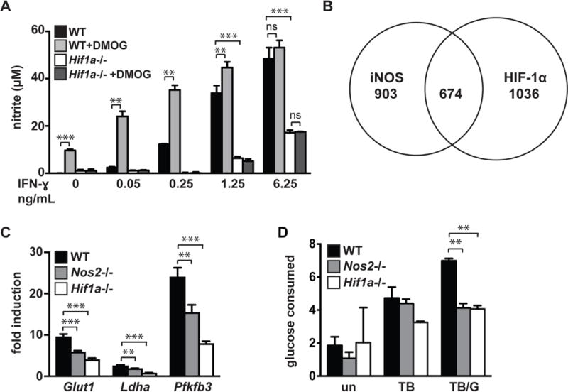
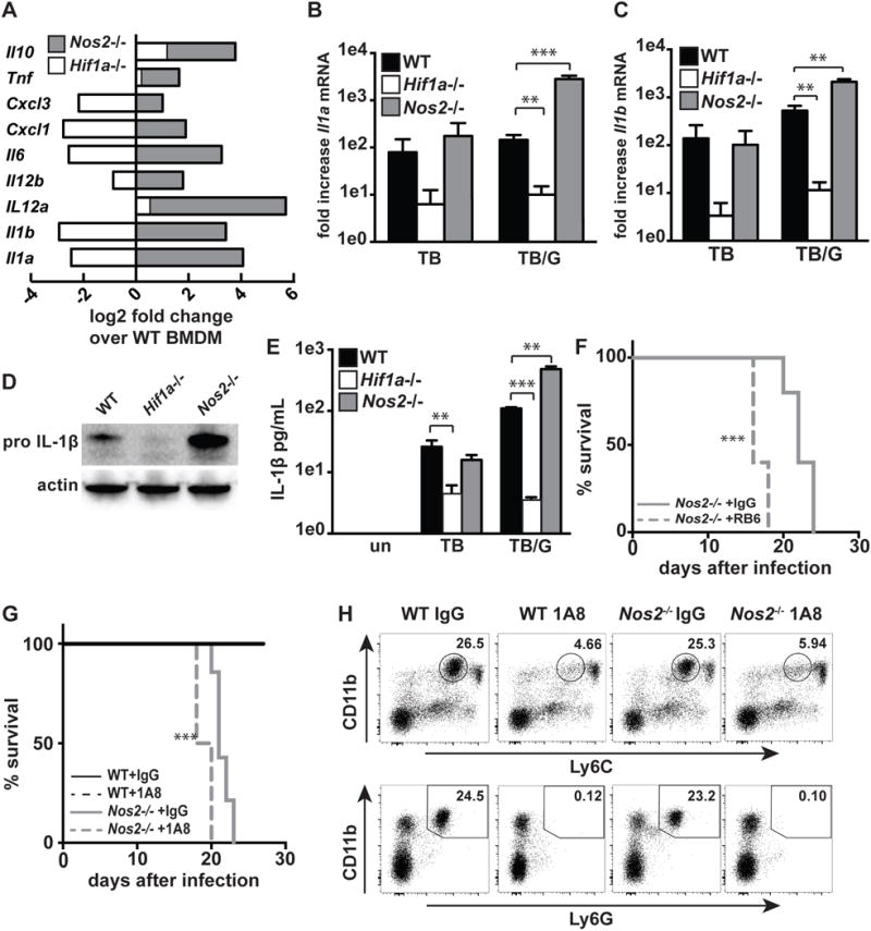
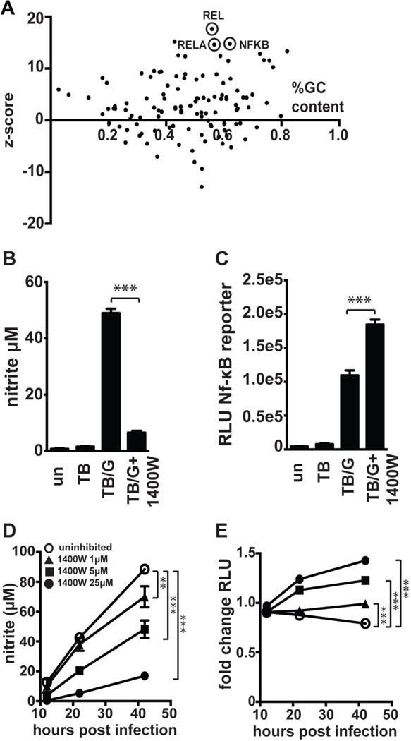
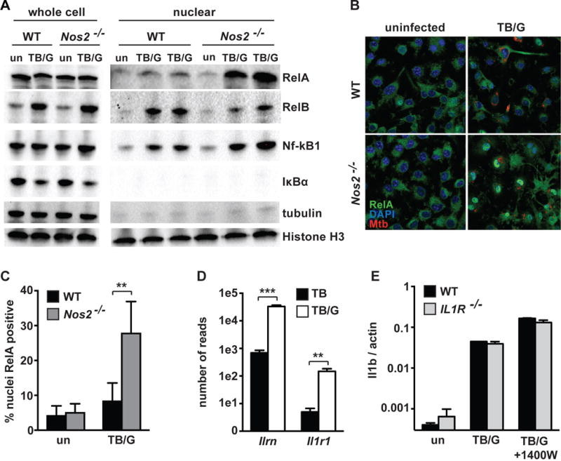
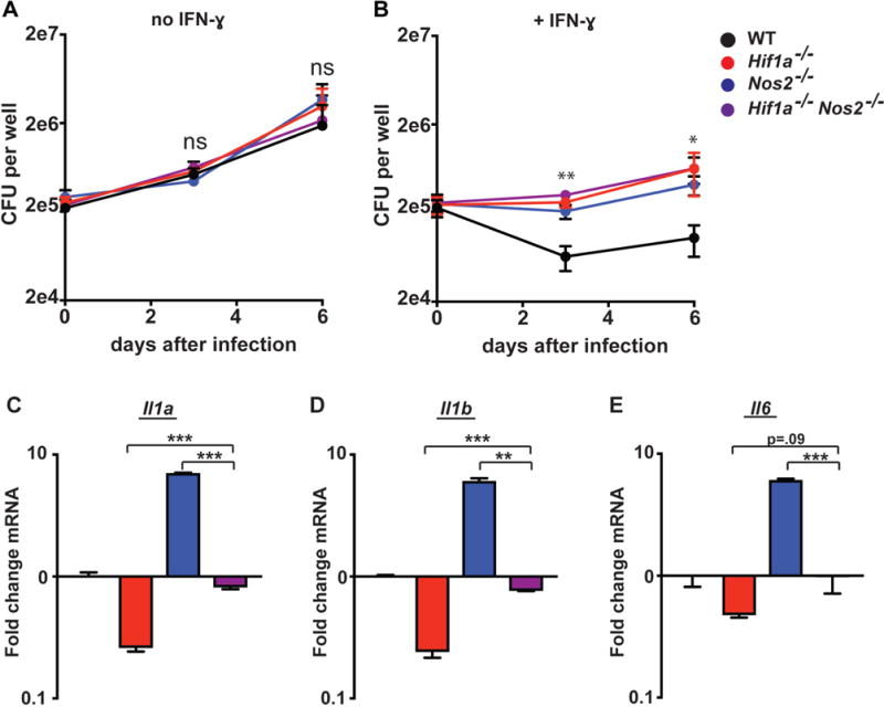
IFN-γ is essential for control of Mycobacterium tuberculosis infection in vitro and in vivo. However, the mechanisms by which IFN-γ controls infection remain only partially understood. One of the crucial IFN-γ target genes required for control of M. tuberculosis is inducible NO synthase (iNOS). Although NO produced by iNOS is thought to have direct bactericidal activity against M. tuberculosis, the role of NO as a signaling molecule has been poorly characterized in the context M. tuberculosis infection. In this study, we found that iNOS broadly regulates the macrophage transcriptome during M. tuberculosis infection, activating antimicrobial pathways while also limiting inflammatory cytokine production. The transcription factor hypoxia inducible factor-1α (HIF-1α) was recently shown to be critical for IFN-γ-mediated control of M. tuberculosis infection. We found that HIF-1α function requires NO production, and that HIF-1α and iNOS are linked by a positive feedback loop that amplifies macrophage activation. Furthermore, we found that NO inhibits NF-κB activity to prevent hyperinflammatory responses. Thus, NO activates robust microbicidal programs while also limiting damaging inflammation. IFN-γ signaling must carefully calibrate an effective immune response that does not cause excessive tissue damage, and this study identifies NO as a key player in establishing this balance during M. tuberculosis infection.


Figures

Figure 1
Figure 1. NO has large effects on the macrophage transcriptome
(A) Total number of differentially expressed genes between wildtype and Nos2/ BMDM in untreated [un], M. tuberculosis infected [TB], and M. tuberculosis infected and IFN-γ treated [TB/G] BMDM 24h post-infection. For this analysis, statistical significance (p<.05) and a 2-fold or greater difference in expression was used as a cutoff. For RNAseq data and all other in vitro infections, the M. tuberculosis strain Erdman was used at an MOI of 5. (B) Griess assay measuring NO production 24h post-infection of wildtype BMDM with and without IFN-γ. (C) Venn diagram showing overlap of iNOS regulated genes during M. tuberculosis infection with IFN-γ treatment, and genes differentially expressed between M. tuberculosis infected wildtype macrophages with and without IFN-γ (D) Bioinformatic prediction (using oPOSSUM) of transcription factors responsible for regulation of all genes found to be expressed at a lower level in Nos2/ BMDM during M. tuberculosis infection with IFN-γ treatment. (E) RNAseq reads 24h post-infection in wildtype, Nos2/, and Hif1a−/− BMDM in untreated [un], M. tuberculosis infected [TB], and M. tuberculosis infected with IFN-γ activation [TB/G] for HIF-1α target genes Egln3 and (F) Bnip3. RNAseq data is from 3 independent infections. (B) is representative of >5 experiments. The p values were determined using an unpaired t test. **p<.01, ***p<.001
Figure 2
Figure 2. iNOS is required for HIF-1α stabilization and transcriptional activity
(A) RNAseq reads for Hif1a 24h post-infection in wildtype and Nos2 BMDM in untreated [un], M. tuberculosis infected [TB], and M. tuberculosis infected with IFN-γ activation [TB/G]. (B) Western blot for HIF-1α in M. tuberculosis infected, IFN-γ activated BMDM at 0,4,12 and 18h post-infection in wildtype and Nos2/ BMDM. Values for quantification of HIF-1α/tubulin ratios are normalized to lane 3 (maximal HIF-1α levels) and are shown below the image. (C) Western blot for HIF-1α during infection with M. tuberculosis with IFN-γ activation in the presence or absence of ascorbate (D) Western blot for HIF-1α in M. tuberculosis infected, IFN-γ activated BMDM with a dose response of the iNOS inhibitor 1400W [.625, 1.25, 2.5, 5, 10, 25uM]. Griess assay for NO production was done on the supernatant of the cells used for the western blot. (E) Western blot for HIF-1α in M. tuberculosis infected, IFN-γ activated BMDM with a dose response of 1400W [.625, 1.25, 2.5, 5uM] with and with addition of the NO donor SNAP [250uM]. (F) HIF-1α levels were measured by western blot at 12h after stimulation with PAM [50ng/ml] and IFN-γ [6.25 ng/ml]. RNAseq data is from 3 independent experiments. All other experiments are representative of 2 or more experiments. Error bars represent the SD and p values were determined using an unpaired t test. ***p<.001
Figure 3
Figure 3. iNOS and HIF-1α are linked by positive feedback, and regulate aerobic glycolysis
(A) Griess assay measuring NO production 24h post-infection of M. tuberculosis infected wildtype and Hif1a−/− BMDM, with a dose response of IFN-γ. The HIF-1α stabilizer DMOG [200uM] increases NO production in a HIF-1α dependent manner. (B) Venn diagram of RNAseq data showing overlap of genes differentially expressed in Nos2/ vs wildtype and Hif1a−/− vs wildtype BMDM during M. tuberculosis infection with IFN-γ activation. (C) RNAseq data showing fold induction of the glycolytic genes Glut1, Ldha, and Pfkfb3 during M. tuberculosis infection with IFN-γ activation in wildtype, Nos2/, Hif1a−/− BMDM relative to untreated. (D) Measurement of glucose uptake in wildtype, Nos2/ and Hif1a−/− BMDM either untreated [un], M. tuberculosis infected [TB], or M. tuberculosis infected and IFN-γ activated [TB/G]. RNAseq data is from 3 independent experiments. (A,D) are representative of 2 or more experiments. Error bars represent SD from 3 or more wells (A,D) or 3 independent experiments (C). The p values were determined using an unpaired t test. **p<.01, ***p<.001
Figure 4
Figure 4. iNOS and HIF-1α antagonistically regulate inflammatory cytokine production
(A) RNAseq data showing fold difference in cytokines and chemokines in Nos2/ and Hif1a−/− BMDM relative to wildtype during M. tuberculosis infection with IFN-γ activation. (B,C) RNAseq data from wildtype, Hif1a−/−, and Nos2/ BMDM showing fold change over uninfected for Il1a and Il1b with either M. tuberculosis infection [TB] or M. tuberculosis infection with IFN-γ activation [TB/G]. (D) Western blot for pro-IL-1B from wildtype, Hif1a−/− and Nos2/ BMDM 24h post-infection with M. tuberculosis and IFN-γ activation. (E) ELISA for IL1B from supernatants of wildtype, Hif1a−/−and Nos2/ BMDM 36h post-infection with and without IFN-γ activation. (F,G) Survival of Nos2/ mice following aerosol infection with the virulent M. tuberculosis strain Erdman, with antibody mediated neutrophil depletion or with control IgG treatment. Mice injected with RB6 (F) or 1A8 (G) antibody every other day beginning 10 days post-infection. (H) Flow cytometry confirming neutrophil depletion from mice treated with 1A8 antibody. Data is from peripheral blood, after 3 antibody injections (15 days post-infection). RNAseq data is from 3 independent experiments. (D,E,F,G) are representative of 2 or more experiments. The p values were determined using an unpaired t test for (B,C,E), and using the Log-rank Mantel-Cox test for (F and G). **p<.01, ***p<.001
Figure 5
Figure 5. iNOS suppresses NF-kB activity
(A) Bioinformatic analysis of RNAseq data using oPOSSUM to predict transcription factors regulating the subset of genes found to be 4-fold or more upregulated in Nos2/ BMDM relative to wildtype BMDM during M. tuberculosis infection with IFN-γ activation. (B) Griess assay measuring NO production 24h post-infection in RAW macrophages carrying an NF-kB luciferase reporter, with M. tuberculosis infection without IFN-γ [TB], with IFN-γ activation [TB/G], and with IFN-γ activation and addition of 25uM 1400W [TB/G+1400W]. (C) Luciferase assay 24h post-infection for the same cells as in (B) to measure NF-kB activity. (D) Griess assays measuring NO production over a timecourse in M. tuberculosis infected, IFN-γ activated BMDM (carrying an NF-kB luciferase reporter) with a dose response of 1400W. (E) Luciferase assay for NF-kB activity from the same cells as in (D). (B,C) representative of 3 experiments, (D,E) representative of 2 experiments. The p values were determined using an unpaired t test. **p<.01, ***p<.001
Figure 6
Figure 6. iNOS deficiency leads to aberrantly high nuclear RelA
(A) Western blots 24h post-infection from whole cell lysates and nuclear extracts for RelA, RelB, NF-kB1, and IkBa, with tubulin as a cytoplasmic marker and Histone H3 as a nuclear marker. Western blots with wildtype and Nos2/ BMDM either uninfected [un] or M. tuberculosis infected with IFN-γ activation [TB/G]. For nuclear extracts, [TB/G] performed in biological duplicate for each repeat. (B) Confocal microscopy at 63x for RelA 24h post-infection in wildtype and Nos2/ BMDM, uninfected or during M. tuberculosis infection with IFN-γ activation [TB/G]. For microscopy, the M. tuberculosis strain Erdman, carrying a fluorescent mCherry was utilized. (C) Quantification of p65 positive nuclei from the same samples as in (B), but from 20x images. >800 nuclei were analyzed for each condition, error bars represent SD between 5 fields. (D) RNAseq data showing number of reads for the IL1R antagonist (Ilrn) and IL1R (Il1r1) in wildtype BMDM infected with M. tuberculosis [TB] or M. tuberculosis with IFN-γ activation [TB/G]. (E) qPCR for Il1b transcript in wildtype and Il1r/ BMDM, either untreated [un], during M. tuberculosis infection with IFN-γ activation [TB/G], or during M. tuberculosis infection with IFN-γ activation and 1400W treatment at 25uM [TB/G+1400W]. All data representative of 2 or more experiments. The p values were determined using an unpaired t test. **p<.01, ***p<.001
Figure 7
Figure 7. Double knockout of iNOS and HIF-1α balances inflammation
Wildtype, Nos2/, Hif1a−/−, and Nos2/ Hif1a−/− BMDM were infected with M. tuberculosis without IFN-γ (A) and with IFN-γ (B). CFU was enumerated immediately following phagocytosis of bacteria, and again 3d and 6d post-infection. (C,D,E) wildtype, Nos2/, Hif1a−/−, and Nos2/ Hif1a−/− BMDM were treated with IFN-γ and infected with M. tuberculosis. qPCR for Il1a, Il1b, and Il6 was performed on RNA isolated from BMDM 24h post-infection. (A–E) representative of 3 or more independent experiments. The p values were determined using an unpaired t test. *p<.05, **p<.01, ***p<.001

References

    1. Floyd K. World Health Organizationed. WHO Press; 2016. pp. 1–100.
    1. Cooper AM, Dalton DK, Stewart TA, Griffin JP, Russell DG, Orme IM. Disseminated tuberculosis in interferon gamma gene-disrupted mice. J Exp Med. 1993;178:2243–2247. - PMC - PubMed
    1. Flynn JL, Chan J, Triebold KJ, Dalton DK, Stewart TA, Bloom BR. An essential role for interferon gamma in resistance to Mycobacterium tuberculosis infection. 1993;178:2249–2254. - PMC - PubMed
    1. Abel L, Casanova J-L. Genetic predisposition to clinical tuberculosis: bridging the gap between simple and complex inheritance. American journal of human genetics. 67:274. - PMC - PubMed
    1. Chan J, Xing Y, Magliozzo RS, Bloom BR. Killing of virulent Mycobacterium tuberculosis by reactive nitrogen intermediates produced by activated murine macrophages. J Exp Med. 175:1111–1122. - PMC - PubMed

Publication types

MeSH terms