. 2017 Jul 28;7(1):6823.
doi: 10.1038/s41598-017-07248-y.

Gyrocardiography: A New Non-invasive Monitoring Method for the Assessment of Cardiac Mechanics and the Estimation of Hemodynamic Variables

Affiliations

Gyrocardiography: A New Non-invasive Monitoring Method for the Assessment of Cardiac Mechanics and the Estimation of Hemodynamic Variables

Mojtaba Jafari Tadi et al. Sci Rep. .

Abstract

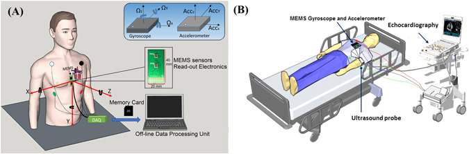
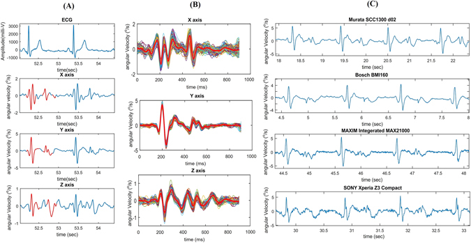
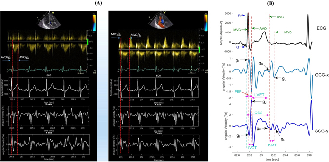
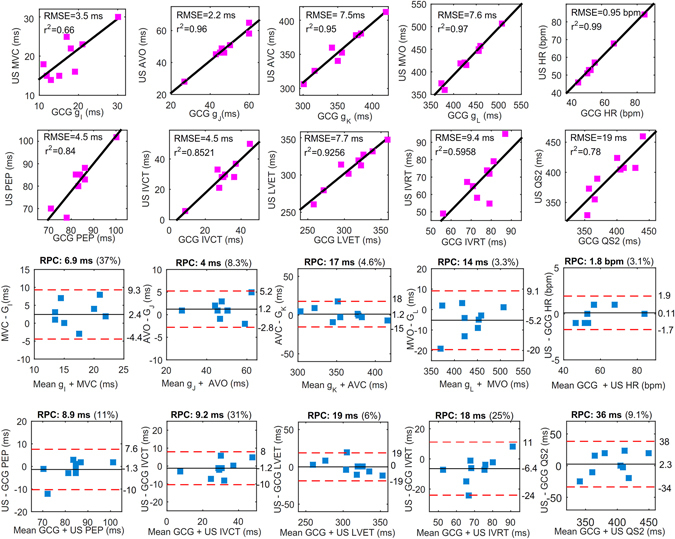
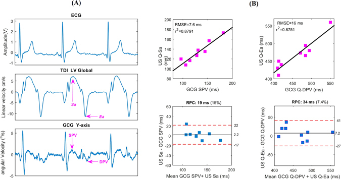
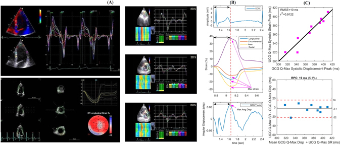
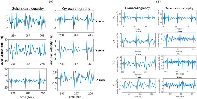
Gyrocardiography (GCG) is a new non-invasive technique for assessing heart motions by using a sensor of angular motion - gyroscope - attached to the skin of the chest. In this study, we conducted simultaneous recordings of electrocardiography (ECG), GCG, and echocardiography in a group of subjects consisting of nine healthy volunteer men. Annotation of underlying fiducial points in GCG is presented and compared to opening and closing points of heart valves measured by a pulse wave Doppler. Comparison between GCG and synchronized tissue Doppler imaging (TDI) data shows that the GCG signal is also capable of providing temporal information on the systolic and early diastolic peak velocities of the myocardium. Furthermore, time intervals from the ECG Q-wave to the maximum of the integrated GCG (angular displacement) signal and maximal myocardial strain curves obtained by 3D speckle tracking are correlated. We see GCG as a promising mechanical cardiac monitoring tool that enables quantification of beat-by-beat dynamics of systolic time intervals (STI) related to hemodynamic variables and myocardial contractility.


Conflict of interest statement

The authors declare that they have no competing interests.

Figures

Figure 1
Figure 1
Simultaneous data acquisition from echocardiography, ECG, and MEMS sensors. General schematics of MEMS motion processing system (A) and echocardiography set up including MEMS sensors and ECG (B).
Figure 2
Figure 2
Typical three dimensional GCG waveforms from x, y, and z axes of rotation. 3-axis GCG morphologies and reference ECG (A). 3-axis ensemble averaged GCG morphologies (B). GCG y-axis waveforms obtained using different sensors (C).
Figure 3
Figure 3
Waveform annotation and cardiac time interval estimation in GCG signal. Aortic (left) and mitral (right) valve opening and closure moments as measured by PW Doppler and correspondingly in GCG signal (A). Waveform annotation in GCG and corresponding time intervals with respect to ECG peaks (B).
Figure 4
Figure 4
Correlation and Bland-Altman plots. The red color dashed lines drawn in Bland-Altman plots represent the upper and lower LoA ranges for the measured cardiac time intervals. RPC is the reproducibility coefficient value which is the maximum difference that is likely to occur between different observations. The coefficient of variation (CV) percentage is the ratio of the standard deviation and the overall mean.
Figure 5
Figure 5
Sa and Ea wave evaluations with TDI and GCG. Qualitative comparison between the TDI Sa and Ea waves and corresponding SPV and DPV in typical GCG y- and z-axis waves (A). Quantitative evaluation of time intervals between Q-Sa/Ea versus Q-SPV/DPV waves (B).
Figure 6
Figure 6
Myocardium tissue velocity, displacement, and strain analysis using TDI and corresponding GCG based angular rates (y-axis). Tissue velocity and displacement using TDI and 3D speckle tracking strain in longitudinal, circumferential and radial directions (A). Electromechanical delays measured by TDI and GCG (B). Relationship between the GCG and TDI electromechanical delays (C).
Figure 7
Figure 7
Visual comparison of GCG and SCG signals. Evaluation of signal quality in typical tri-axial SCG and GCG waveforms (A). Intersubject variability comparison for GCG against SCG (B).

References

    1. Waldman LK, Fung YC, Covell JW. Transmural myocardial deformation in the canine left ventricle. Normal in vivo three-dimensional finite strains. Circulation Research. 1985;57:152–63. doi: 10.1161/01.RES.57.1.152. - DOI - PubMed
    1. Rademakers FE, et al. Dissociation between left ventricular untwisting and filling. Accentuation by catecholamines. Circulation. 1992;85:1572–81. doi: 10.1161/01.CIR.85.4.1572. - DOI - PubMed
    1. Croone, W. & Maquet, P. On the reason of the movement of the muscles. Transactions of the American Philosophical Society (American Philosophical Society, 2000).
    1. Taber LA, Yang M, Podszus W. Mechanics of ventricular torsion. Journal of Biomechanics. 1996;29:745–752. doi: 10.1016/0021-9290(95)00129-8. - DOI - PubMed
    1. Ingels NB, Daughters GT, Stinson EB, Alderman EL. Measurement of midwall myocardial dynamics in intact man by radiography of surgically implanted markers. Circulation. 1975;52:859–67. doi: 10.1161/01.CIR.52.5.859. - DOI - PubMed

Publication types