. 2017 Jul 28;7(1):6787.
doi: 10.1038/s41598-017-07010-4.

High-throughput mutagenesis using a two-fragment PCR approach

Affiliations

High-throughput mutagenesis using a two-fragment PCR approach

Franziska M Heydenreich et al. Sci Rep. .

Abstract

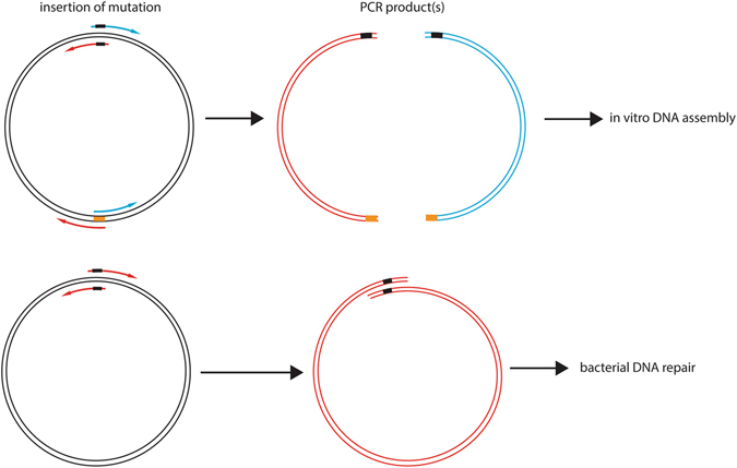
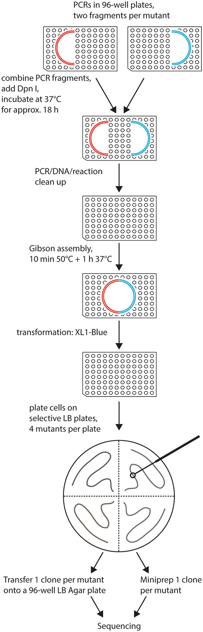
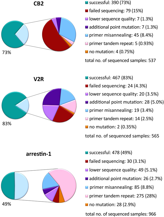
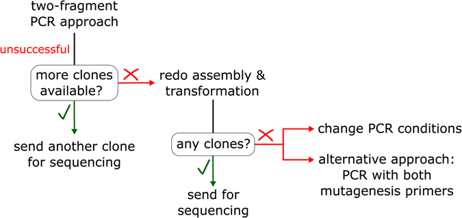
Site-directed scanning mutagenesis is a powerful protein engineering technique which allows studies of protein functionality at single amino acid resolution and design of stabilized proteins for structural and biophysical work. However, creating libraries of hundreds of mutants remains a challenging, expensive and time-consuming process. The efficiency of the mutagenesis step is the key for fast and economical generation of such libraries. PCR artefacts such as misannealing and tandem primer repeats are often observed in mutagenesis cloning and reduce the efficiency of mutagenesis. Here we present a high-throughput mutagenesis pipeline based on established methods that significantly reduces PCR artefacts. We combined a two-fragment PCR approach, in which mutagenesis primers are used in two separate PCR reactions, with an in vitro assembly of resulting fragments. We show that this approach, despite being more laborious, is a very efficient pipeline for the creation of large libraries of mutants.


Conflict of interest statement

The authors declare that they have no competing interests.

Figures

Figure 1
Figure 1
Comparison of mutagenesis techniques. In the two-fragment approach, the two mutagenesis primers are separated into two PCR reactions and combined with one reverse primer on the opposite side of the vector respectively, while the one-fragment approach relies on two mutagenesis primers in one PCR reaction.
Figure 2
Figure 2
Agarose gel electrophoresis analysis of PCR fragments multiplied by Phusion High-Fidelity PCR Master Mix with HF Buffer and Phusion High-Fidelity PCR Master Mix with GC Buffer. Analysed PCRs are labelled as the mutated residue and the letter indicating whether the fragment is multiplied by mutation-specific forward (F) or reverse (R) primer. Expected PCR fragments are between 3000 and 4000 bp long. While using the PCR master mix with GC buffer gave expected fragments in all shown cases, using the Phusion High-Fidelity polymerase with HF buffer failed to multiply four fragments.
Figure 3
Figure 3
Overview of the mutagenesis technique. Two PCR reactions are done per mutant, in each of them approximately half of the vector is amplified. Two fragments containing one mutation are combined, followed by DpnI digestion at 37 °C overnight. Reaction clean-up is performed to purify DNA fragments, which are then assembled by Gibson assembly reaction. Bacteria are transformed with the resulting circular plasmid and plated on selective LB agar plates. One clone per mutant is sent for sequencing either on a selective LB agar 96-well plate or as purified DNA. All steps excluding the plating of the bacteria are done in 96-well plates.
Figure 4
Figure 4
Results of alanine scanning mutagenesis on two G protein-coupled receptors, cannabinoid CB2 receptor (CB2) and vasopressin V2 receptor (V2R) compared with the one-fragment mutagenesis approach used for arrestin-1 (Sun, Ostermaier et al.). Reason for failure and corresponding percentage within the total number of DNA samples sent for sequencing are given. In case of V2R and arrestin-1, the total number of analyzed samples is smaller than the sum of individual categories as in a few instances several failure reasons could be found within a sequenced sample. Category “failed sequencing” relates to instances with very noisy peaks or no peaks at all in a sequencing electrophoretogram. On the other hand, “lower sequence quality” denotes interpretable sequence traces that had some artefacts or were too short to provide reliable information about success of mutagenesis.
Figure 5
Figure 5
Troubleshooting scheme. For mutants which were not obtained in the first round of cloning, another clone can be sequenced, if it exists. Furthermore, the Gibson assembly reaction can be redone with the same purified DNA fragments, followed by bacterial transformation and sequencing. For missing mutants, PCR conditions can be changed or PCR containing both mutagenesis primers (one-fragment approach) can be applied.
Figure 6
Figure 6
GC content of the plasmids used for the generation of the mutagenesis libraries. For each nucleotide position in the plasmid sequence, GC content was calculated using a 25 bp sliding window. The mutated sequences are highlighted in red.

References

    1. Duncan AR, Winter G. The binding site for C1q on IgG. Nature. 1988;332:738–740. doi: 10.1038/332738a0. - DOI - PubMed
    1. Cunningham, B. C. & Wells, J. A. High-resolution epitope mapping of hGH-receptor interactions by alanine-scanning mutagenesis. Science244, 1081–1085, doi:10.1126/science.2471267 (1989). - PubMed
    1. Heydenreich FM, Vuckovic Z, Matkovic M, Veprintsev DB. Stabilization of G protein-coupled receptors by point mutations. Front Pharmacol. 2015;6:82. doi: 10.3389/fphar.2015.00082. - DOI - PMC - PubMed
    1. Ostermaier MK, Peterhans C, Jaussi R, Deupi X, Standfuss J. Functional map of arrestin-1 at single amino acid resolution. Proc Natl Acad Sci USA. 2014;111:1825–1830. doi: 10.1073/pnas.1319402111. - DOI - PMC - PubMed
    1. Sun D, et al. Probing Galphai1 protein activation at single-amino acid resolution. Nat Struct Mol Biol. 2015;22:686–694. doi: 10.1038/nsmb.3070. - DOI - PMC - PubMed

Publication types

MeSH terms