

A Modified Preserved Nasal and Lacrimal Flap Technique in Endoscopic Dacryocystorhinostomy
- PMID: 28754905
- PMCID: PMC5533767
- DOI: 10.1038/s41598-017-07364-9
A Modified Preserved Nasal and Lacrimal Flap Technique in Endoscopic Dacryocystorhinostomy
Abstract
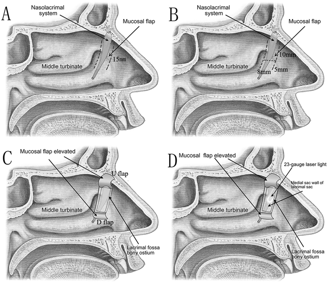
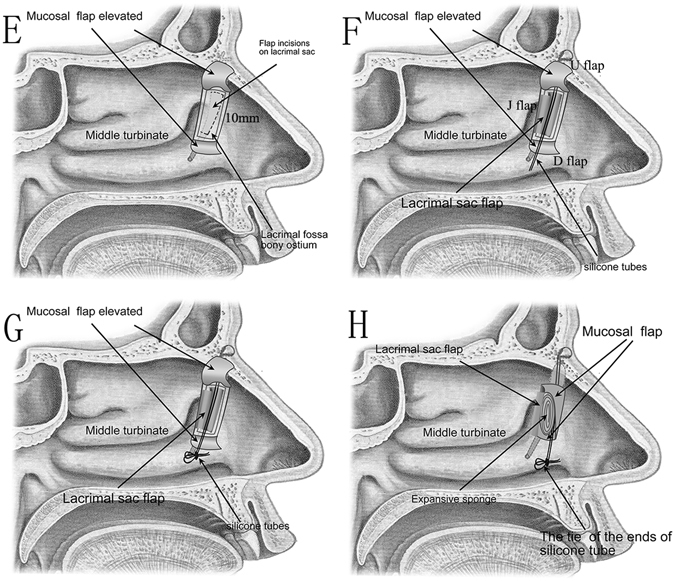
Here we describe a modified preserved nasal and lacrimal mucosal flap technique in endonasal endoscopic dacryocystorhinostomy (EES-DCR) for patients with epiphora secondary to primary acquired nasolacrimal duct obstruction (PANDO) and evaluate its outcomes. Twenty-five patients with PANDO were retrospectively reviewed. Modified preserved nasal and lacrimal mucosal flap technique in EES-DCR was applied in all 27 eyes of 25 patients. The patients were evaluated with objective (anatomical patency) and subjective (symptomatic cure) success rates within the duration of follow-up. In the present study, all of the patients' surgical procedures were successful. There were 2 cases of flap dislocation from the rhinostomy site 1 week post-operation. After a mean follow-up of 4.9 ± 1.8 months, the success rate of anatomical patency was 100% (27/27) and the success rate of symptomatic cure was 92.6% (25/27). No significant complications occurred intraoperatively. We concluded that the modified preserved nasal and lacrimal mucosal flap technique in EES-DCR for treating PANDO is simple and safe, can effectively cover the bare bone around the opened sac, and provide a similar or even better clinical outcome compared with other routine treatment techniques used for this condition.
Conflict of interest statement
The authors declare that they have no competing interests.
Figures
Similar articles
-
Endonasal dacryocystorhinostomy: a modified technique with preservation of the nasal and lacrimal mucosa.Ophthalmic Plast Reconstr Surg. 2010 May-Jun;26(3):161-4. doi: 10.1097/IOP.0b013e3181b80af6. Ophthalmic Plast Reconstr Surg. 2010. PMID: 20489538
-
Prevention of re-obstruction in watery eye treatment: three-flap technique in external dacryocystorhinostomy.Graefes Arch Clin Exp Ophthalmol. 2016 Dec;254(12):2455-2460. doi: 10.1007/s00417-016-3490-z. Epub 2016 Sep 2. Graefes Arch Clin Exp Ophthalmol. 2016. PMID: 27590057
-
Comparison of surgical outcomes of endonasal dacryocystorhinostomy with or without mucosal flaps.Auris Nasus Larynx. 2009 Oct;36(5):555-9. doi: 10.1016/j.anl.2009.01.005. Epub 2009 Mar 17. Auris Nasus Larynx. 2009. PMID: 19297108
-
Evidence-based review of surgical practices in endoscopic endonasal dacryocystorhinostomy for primary acquired nasolacrimal duct obstruction and other new indications.Curr Opin Ophthalmol. 2014 Sep;25(5):443-8. doi: 10.1097/ICU.0000000000000084. Curr Opin Ophthalmol. 2014. PMID: 24979582 Review.
-
Influence of Surgical Techniques on Endoscopic Dacryocystorhinostomy: A Systematic Review and Meta-analysis.Otolaryngol Head Neck Surg. 2021 Jul;165(1):14-22. doi: 10.1177/0194599820972677. Epub 2020 Nov 24. Otolaryngol Head Neck Surg. 2021. PMID: 33228432
Cited by
-
Nasolacrimal duct rhinostomy for low-level nasolacrimal duct obstruction:long-term outcomes and surgical selection paradigm.Eur Arch Otorhinolaryngol. 2024 Nov;281(11):5783-5792. doi: 10.1007/s00405-024-08797-5. Epub 2024 Aug 6. Eur Arch Otorhinolaryngol. 2024. PMID: 39107549
-
A Comparative Study Between Anterior-Posterior and Superior-Inferior Flap Suturing Technique in Endoscopic Dacryocystorhinostomy at our Tertiary Institution.Indian J Otolaryngol Head Neck Surg. 2023 Dec;75(4):2927-2935. doi: 10.1007/s12070-023-03860-9. Epub 2023 May 26. Indian J Otolaryngol Head Neck Surg. 2023. PMID: 37974788 Free PMC article.
-
Endoscopic endonasal dacryocystorhinostomy learning curve.Arq Bras Oftalmol. 2022 May-Jun;85(3):223-228. doi: 10.5935/0004-2749.20220030. Arq Bras Oftalmol. 2022. PMID: 34586231 Free PMC article.
-
[Effect of endonasal endoscopic dacryocystorhinostomy combined with lacrimal duct drainage tube implantation in treating lacrimal duct obstruction].Lin Chuang Er Bi Yan Hou Tou Jing Wai Ke Za Zhi. 2022 Nov;36(11):845-848. doi: 10.13201/j.issn.2096-7993.2022.11.007. Lin Chuang Er Bi Yan Hou Tou Jing Wai Ke Za Zhi. 2022. PMID: 36347577 Free PMC article. Chinese.
-
Comparative Study of Endonasal Endoscopic Dacryocystorhinostomy with or without Preservation of Nasal Mucosal Flap.Indian J Otolaryngol Head Neck Surg. 2024 Feb;76(1):894-898. doi: 10.1007/s12070-023-04311-1. Epub 2023 Oct 31. Indian J Otolaryngol Head Neck Surg. 2024. PMID: 38440443 Free PMC article.
References
-
- Toti A. Nuovo metodo conservatore di cura radicale delle suppurazioni croniche del sacco lacrimale (dacriocistorinostomia) Clin Mod Firenze. 1904;10:385–387.
-
- Zilelioglu G, et al. Results of endoscopic endonasal non-laser dacryocystorhinostomy. Documenta ophthalmologica. Advances in ophthalmology. 2002;105:57–62. - PubMed
Publication types
MeSH terms
LinkOut - more resources
Full Text Sources
Other Literature Sources
Medical
