

KRAS Activation and over-expression of SIRT1/BCL6 Contributes to the Pathogenesis of Endometriosis and Progesterone Resistance
- PMID: 28754906
- PMCID: PMC5533722
- DOI: 10.1038/s41598-017-04577-w
KRAS Activation and over-expression of SIRT1/BCL6 Contributes to the Pathogenesis of Endometriosis and Progesterone Resistance
Abstract
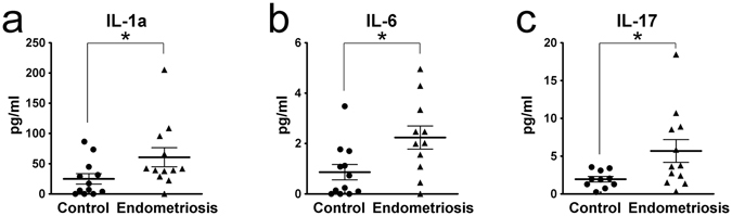
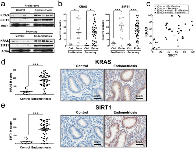
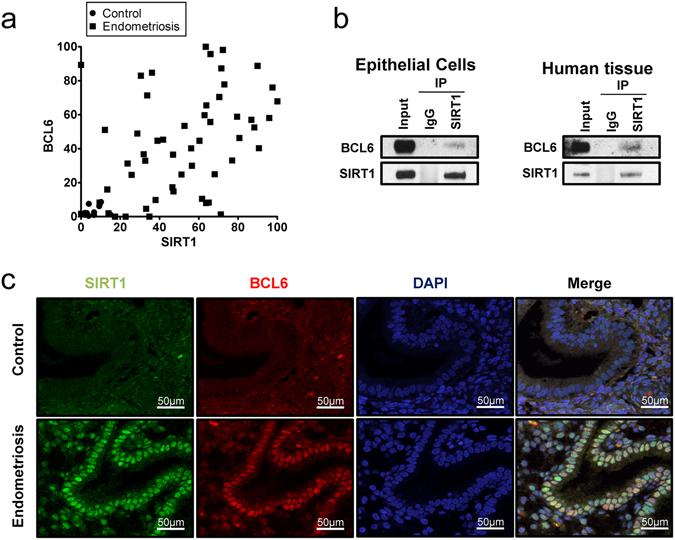
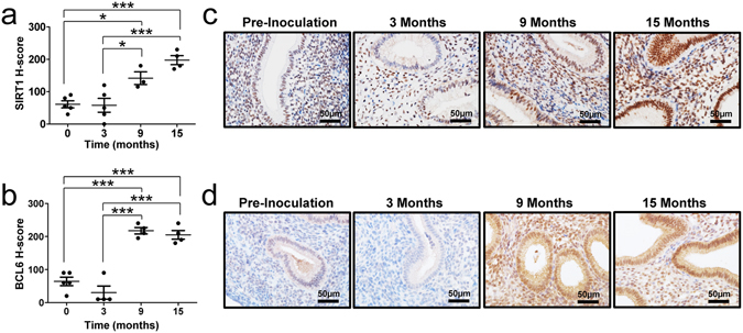
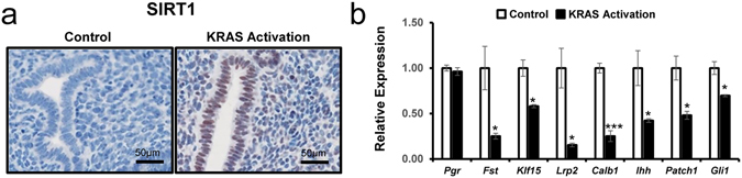
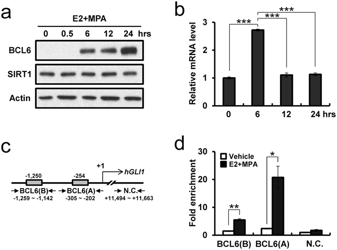
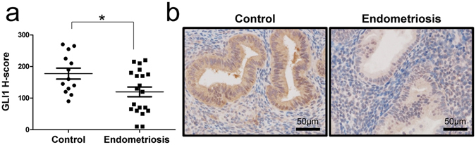
Endometriosis is an inflammatory condition that is associated with progesterone resistance and cell proliferation, resulting in pain, infertility and pregnancy loss. We previously demonstrated phosphorylation of STAT3 in eutopic endometrium of infertile women with this disorder leading to over-expression of the oncogene BCL6 and stabilization of hypoxia-induced factor 1 alpha (HIF-1α). Here we report coordinated activation of KRAS and over-expression of Sirtuin 1 (SIRT1), a histone deacetylase and gene silencer, in the eutopic endometrium from women with endometriosis throughout the menstrual cycle. The mice with conditional activation of KRAS in the PGR positive cells reveal an increase of SIRT1 expression in the endometrium compared to control mice. The expression of progesterone receptor target genes including the Indian Hedgehog pathway genes are significantly down-regulated in the mutant mice. SIRT1 co-localizes with BCL6 in the nuclei of affected individuals and both proteins bind to and suppress the promoter of GLI1, a critical mediator of progesterone action in the Indian Hedgehog pathway, by ChIP analysis. In eutopic endometrium, GLI1 expression is reduced in women with endometriosis. Together, these data suggest that KRAS, SIRT1 and BCL6 are coordinately over-expressed in eutopic endometrium of women with endometriosis and likely participate in the pathogenesis of endometriosis.
Conflict of interest statement
The authors declare that they have no competing interests.
Figures
Similar articles
-
Role of SIRT1 and Progesterone Resistance in Normal and Abnormal Endometrium.J Clin Endocrinol Metab. 2022 Feb 17;107(3):788-800. doi: 10.1210/clinem/dgab753. J Clin Endocrinol Metab. 2022. PMID: 34665857 Free PMC article.
-
Differential Expression of KRAS and SIRT1 in Ovarian Cancers with and Without Endometriosis.Reprod Sci. 2020 Jan;27(1):145-151. doi: 10.1007/s43032-019-00017-4. Epub 2020 Jan 1. Reprod Sci. 2020. PMID: 32046380
-
miR-196a overexpression activates the MEK/ERK signal and represses the progesterone receptor and decidualization in eutopic endometrium from women with endometriosis.Hum Reprod. 2016 Nov;31(11):2598-2608. doi: 10.1093/humrep/dew223. Epub 2016 Sep 12. Hum Reprod. 2016. PMID: 27619769
-
Progesterone Resistance in Endometriosis: an Acquired Property?Trends Endocrinol Metab. 2018 Aug;29(8):535-548. doi: 10.1016/j.tem.2018.05.006. Epub 2018 Jun 19. Trends Endocrinol Metab. 2018. PMID: 29934050 Review.
-
Adenomyosis pathogenesis: insights from next-generation sequencing.Hum Reprod Update. 2021 Oct 18;27(6):1086-1097. doi: 10.1093/humupd/dmab017. Hum Reprod Update. 2021. PMID: 34131719 Free PMC article. Review.
Cited by
-
Discovery of 5-Benzylidene-2-phenyl-1,3-dioxane-4,6-diones as Highly Potent and Selective SIRT1 Inhibitors.ACS Med Chem Lett. 2021 Mar 1;12(3):397-403. doi: 10.1021/acsmedchemlett.0c00559. eCollection 2021 Mar 11. ACS Med Chem Lett. 2021. PMID: 33738067 Free PMC article.
-
Non-Invasive and Mechanism-Based Molecular Assessment of Endometrial Receptivity During the Window of Implantation: Current Concepts and Future Prospective Testing Directions.Front Reprod Health. 2022 May 4;4:863173. doi: 10.3389/frph.2022.863173. eCollection 2022. Front Reprod Health. 2022. PMID: 36303672 Free PMC article. Review.
-
The Origin and Pathogenesis of Endometriosis.Annu Rev Pathol. 2020 Jan 24;15:71-95. doi: 10.1146/annurev-pathmechdis-012419-032654. Epub 2019 Sep 3. Annu Rev Pathol. 2020. PMID: 31479615 Free PMC article. Review.
-
Medical or surgical treatment before embryo transfer improves outcomes in women with abnormal endometrial BCL6 expression.J Assist Reprod Genet. 2019 Mar;36(3):483-490. doi: 10.1007/s10815-018-1388-x. Epub 2019 Jan 4. J Assist Reprod Genet. 2019. PMID: 30610661 Free PMC article.
-
Evaluation of BCL6 and SIRT1 as Non-Invasive Diagnostic Markers of Endometriosis.Curr Issues Mol Biol. 2021 Sep 28;43(3):1350-1360. doi: 10.3390/cimb43030096. Curr Issues Mol Biol. 2021. PMID: 34698105 Free PMC article.
References
Publication types
MeSH terms
Substances
Supplementary concepts
Grants and funding
LinkOut - more resources
Full Text Sources
Other Literature Sources
Medical
Molecular Biology Databases
Research Materials
Miscellaneous
