. 2017 Jul 28;7(1):6835.
doi: 10.1038/s41598-017-07047-5.

In vivo Raman spectral analysis of impaired cervical remodeling in a mouse model of delayed parturition

Affiliations

In vivo Raman spectral analysis of impaired cervical remodeling in a mouse model of delayed parturition

Christine M O'Brien et al. Sci Rep. .

Abstract

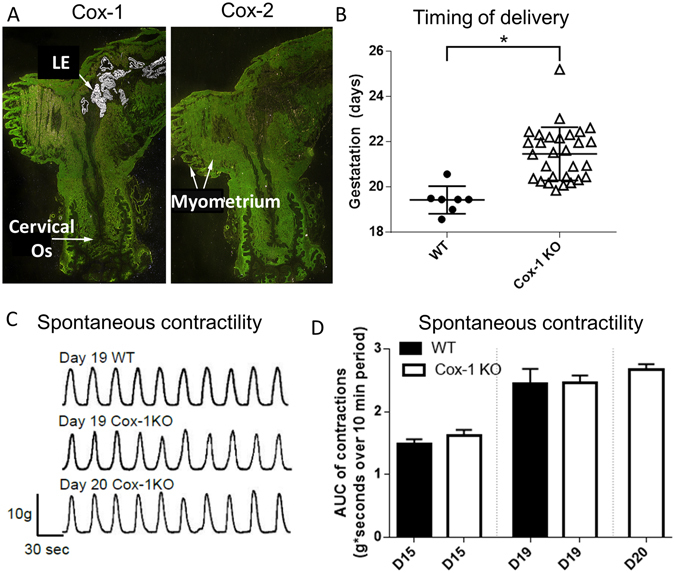
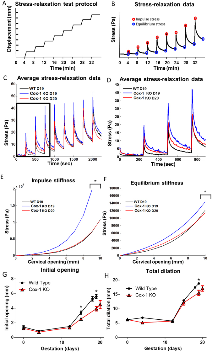
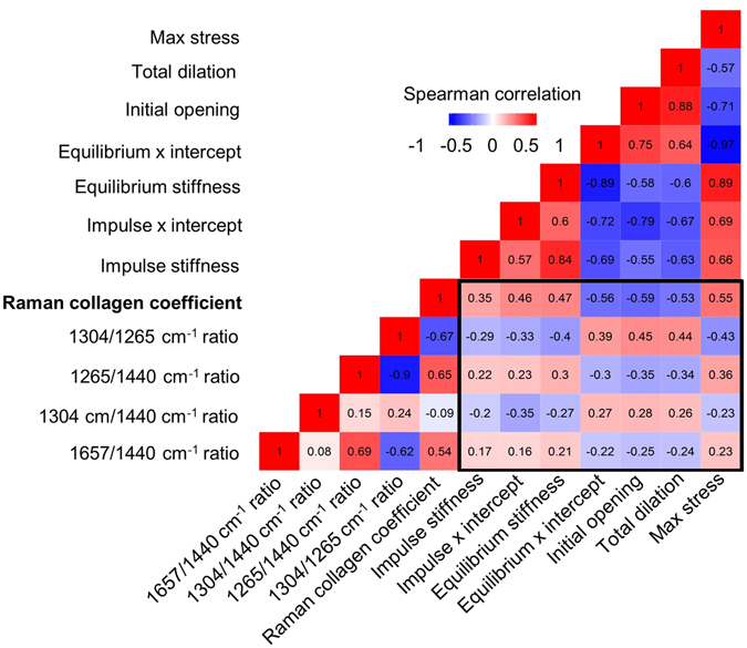
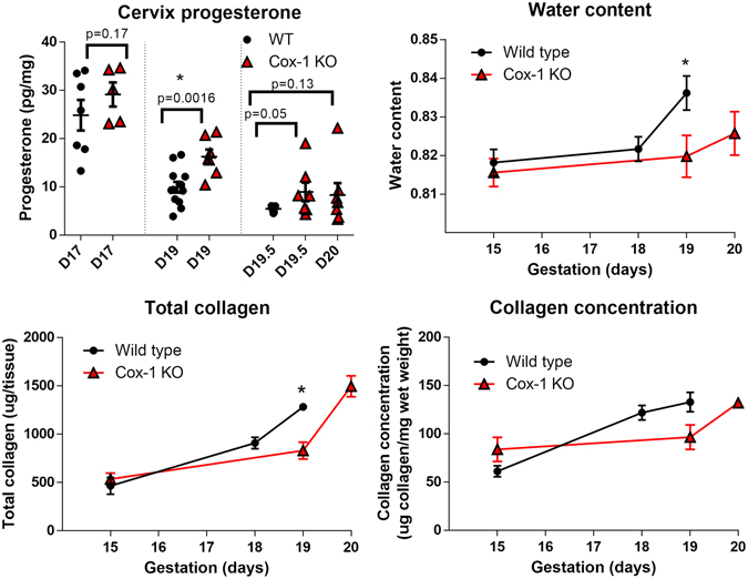
Monitoring cervical structure and composition during pregnancy has high potential for prediction of preterm birth (PTB), a problem affecting 15 million newborns annually. We use in vivo Raman spectroscopy, a label-free, light-based method that provides a molecular fingerprint to non-invasively investigate normal and impaired cervical remodeling. Prostaglandins stimulate uterine contractions and are clinically used for cervical ripening during pregnancy. Deletion of cyclooxygenase-1 (Cox-1), an enzyme involved in production of these prostaglandins, results in delayed parturition in mice. Contrary to expectation, Cox-1 null mice displayed normal uterine contractility; therefore, this study sought to determine whether cervical changes could explain the parturition differences in Cox-1 null mice and gestation-matched wild type (WT) controls. Raman spectral changes related to extracellular matrix proteins, lipids, and nucleic acids were tracked over pregnancy and found to be significantly delayed in Cox-1 null mice at term. A cervical basis for the parturition delay was confirmed by other ex vivo tests including decreased tissue distensibility, hydration, and elevated progesterone levels in the Cox-1 null mice at term. In conclusion, in vivo Raman spectroscopy non-invasively detected abnormal remodeling in the Cox-1 null mouse, and clearly demonstrated that the cervix plays a key role in their delayed parturition.


Conflict of interest statement

The authors declare that they have no competing interests.

Figures

Figure 1
Figure 1
Cox-1 expression in the pregnant mouse uterus, and the effect of global Cox-1 deletion on parturition and uterine contractility. (A) In situ hybridization of 35S-labeled Cox-1 and Cox-2 in WT day 19 mouse uterus. (B) Timing of delivery was recorded from pregnant WT and Cox-1 KO mice (*p = 0.0001). (C) Representative ex vivo contractility tracings of myometrial strips from pregnant mice on the indicated days of pregnancy. (D) Recordings of myometrial contractility (n = 5–11 mice per group) were analyzed for area under the curve (AUC). Mean ± SEM.
Figure 2
Figure 2
In vivo Raman spectroscopy system for cervical assessment during pregnancy. (A) Illustration showing the placement of the Raman spectroscopy fiber optic probe against the ectocervix of the mouse. (B) Mouse cervix visualized using a speculum. (C) In vivo Raman spectroscopy during measurement of mouse cervix. (D) Raman spectroscopy fiber optic probe. (E) Image of in vivo Raman spectroscopy system. Average Raman spectra from different time points during pregnancy in WT (F) and Cox-1 KO (G) mice. Gray boxes indicate regions that were highlighted in Figs 3–4.
Figure 3
Figure 3
Raman spectral bands change with pregnancy and are delayed in the Cox-1 KO mouse compared to WT. Average Raman spectra from the 1265 cm−1 peak (blue dashed line) and 1304 cm−1 peak (blue dashed line) during different time points of pregnancy in WT (A) and Cox-1 KO (B) mice. (C) Mean ± SEM of the 1304 cm−1 to 1265 cm−1 peak ratio as a function of gestation in WT and Cox-1 KO mice (p < 0.05). (D) Modeled longitudinal trajectories of the 1304 cm−1 to 1265 cm−1 Raman peak ratio.
Figure 4
Figure 4
Raman spectra reveal delayed remodeling in the Cox-1 KO mouse at term. Mean ± SEM (A) and modeled longitudinal trajectories (B) of the 1657 cm−1 to 1440 cm−1 peak ratio. Mean ± SEM (C) and modeled longitudinal trajectory (D) of the non-negative least squares model collagen coefficient as a function of gestation in WT and Cox-1 KO mice.
Figure 5
Figure 5
Cox-1 KO mice have less distensible cervices at term than WT. (A) Displacement protocol used for biomechanical stress relaxation tests. (B) Representative data from stress-relaxation biomechanical tests. Red circles highlight the maximum impulse stress observed for each displacement; blue circles highlight the equilibrium stress observed four minutes post displacement. (C) Average stress-relaxation recordings from WT day 19, Cox-1 KO day 19, and Cox-1 day 20 cervical tissues. (D) Inset of the average stress-relaxation data from the first three displacements. (E,F) Average exponential fits to the impulse stiffness and equilibrium stiffness results from WT day 19, Cox-1 KO day 19, and Cox-1 KO day 20 cervical tissues, *p < 0.05 compared to day 19 WT. (G) Unloaded dilation of ex vivo cervical tissues from WT and Cox-1 KO mice across gestation (n = 8–10 per group, per time point). (H) Total dilation of ex vivo cervical tissues prior to tissue failure.
Figure 6
Figure 6
In vivo Raman data correlate with ex vivo biomechanical properties. Spearman correlation matrix containing four Raman peak ratios and a non-negative least squares component representative of collagen signatures for comparison to the impulse stiffness, impulse x intercept, equilibrium stiffness, equilibrium x intercept, initial opening, total dilation, and maximum stress measures obtained during biomechanical testing.
Figure 7
Figure 7
Ex vivo biochemical assays of cervix composition show delayed remodeling and Cox-1 KO mice. (A) Cervix progesterone levels. (B) Water content. (C) Total collagen. (D) Collagen concentration (μg/mg wet weight). Plotted as mean ± SEM, *p < 0.05 compared to day 19 WT.

References

    1. Romero R, Dey SK, Fisher SJ. Preterm Labor: One Syndrome, Many Causes. Science (New York, N.Y) 2014;345:760–765. doi: 10.1126/science.1251816. - DOI - PMC - PubMed
    1. Feltovich, H., Hall, T. & Berghella, V. Beyond cervical length: emerging technologies for assessing the pregnant cervix. Am J of Obstet and Gynecol07 (2012). - PMC - PubMed
    1. Word RA, Li XH, Hnat M, Carrick K. Dynamics of cervical remodeling during pregnancy and parturition: mechanisms and current concepts. Semin Reprod Med. 2007;25:69–79. doi: 10.1055/s-2006-956777. - DOI - PubMed
    1. Yoshida K, et al. Quantitative Evaluation of Collagen Crosslinks and Corresponding Tensile Mechanical Properties in Mouse Cervical Tissue during Normal Pregnancy. PloS one. 2014;9:e112391. doi: 10.1371/journal.pone.0112391. - DOI - PMC - PubMed
    1. Akgul Y, Holt R, Mummert M, Word A, Mahendroo M. Dynamic changes in cervical glycosaminoglycan composition during normal pregnancy and preterm birth. Endocrinology. 2012;153:3493–3503. doi: 10.1210/en.2011-1950. - DOI - PMC - PubMed

Publication types