. 2017 Jul 28;7(1):6812.
doi: 10.1038/s41598-017-07215-7.

Zinc binding to RNA recognition motif of TDP-43 induces the formation of amyloid-like aggregates

Affiliations

Zinc binding to RNA recognition motif of TDP-43 induces the formation of amyloid-like aggregates

Cyrille Garnier et al. Sci Rep. .

Abstract

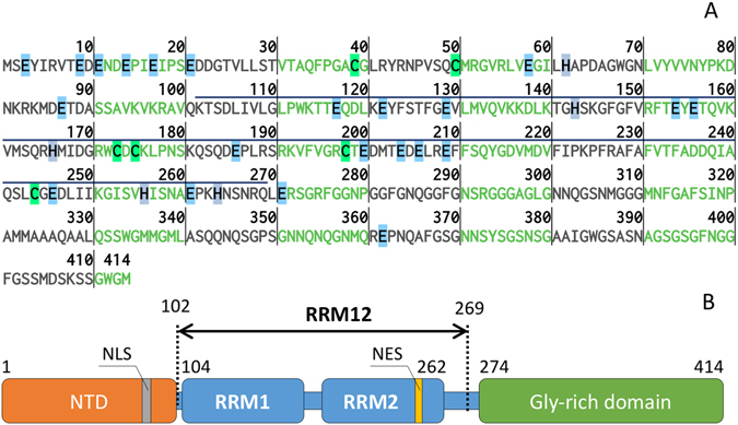
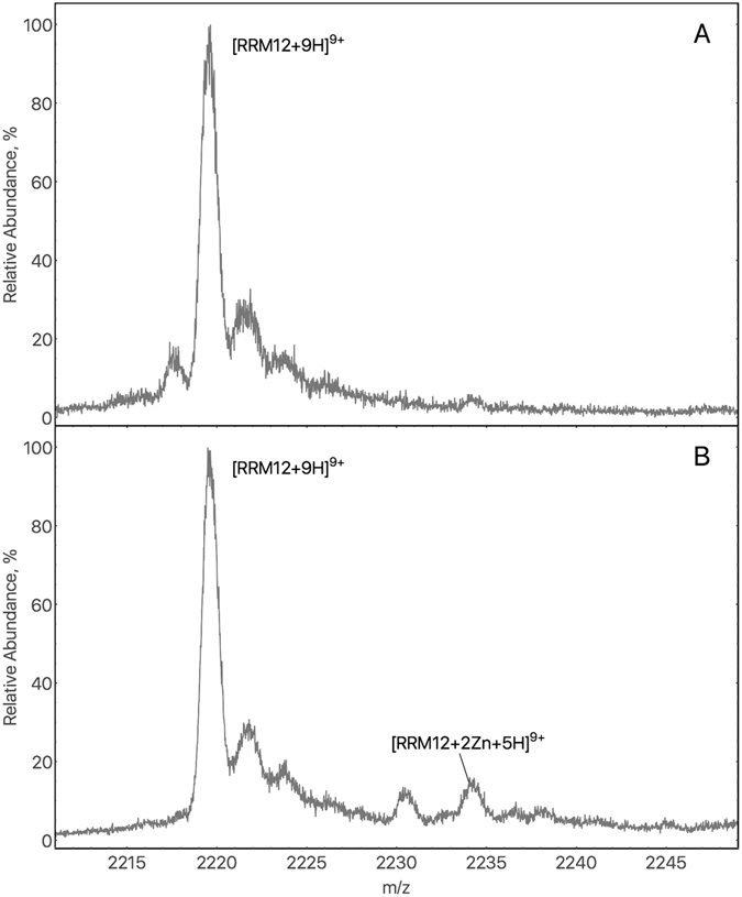
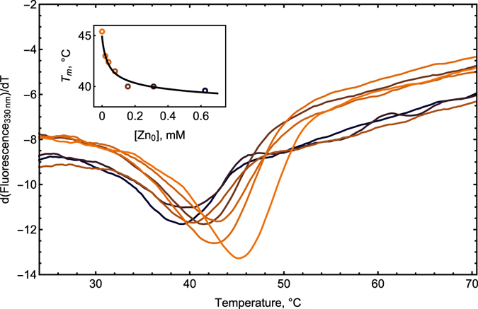
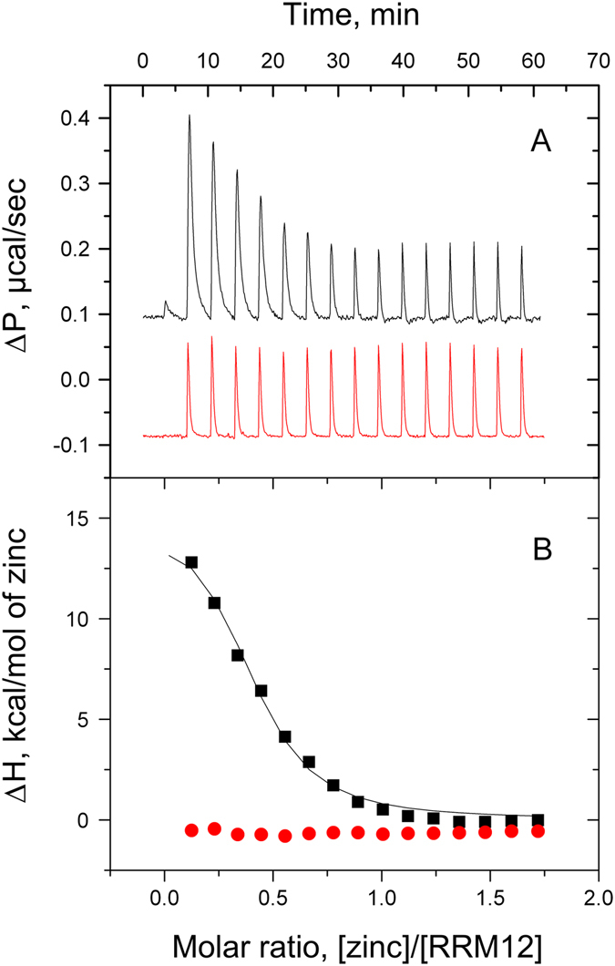
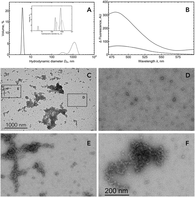
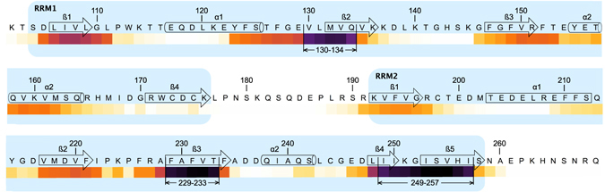
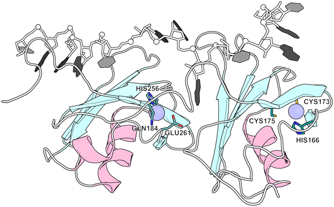
Aggregation of TDP-43 (transactive response DNA binding protein 43 kDa) is a hallmark of certain forms of amyotrophic lateral sclerosis (ALS) and frontotemporal lobar degeneration (FTLD). Moreover, intracellular TDP-43-positive inclusions are often found in other neurodegenerative diseases. Recently it was shown that zinc ions can provoke the aggregation of endogenous TDP-43 in cells, allowing to assume a direct interaction of TDP-43 with zinc ions. In this work, we investigated zinc binding to the 102-269 TDP-43 fragment, which comprise the two RNA recognition motifs. Using isothermal titration calorimetry, mass spectrometry, and differential scanning fluorimetry, we showed that zinc binds to this TDP-43 domain with a dissociation constant in the micromolar range and modifies its tertiary structure leading to a decrease of its thermostability. Moreover, the study by dynamic light scattering and negative stain electron microscopy demonstrated that zinc ions induce auto-association process of this TDP-43 fragment into rope-like structures. These structures are thioflavin-T-positive allowing to hypothesize the direct implication of zinc ions in pathological aggregation of TDP-43.


Conflict of interest statement

The authors declare that they have no competing interests.

Figures

Figure 1
Figure 1
(A) Amino acid sequence of Transactive response DNA-binding protein 43 (TDP-43). The fragment of 102–269 (RRM12) used in this study is overlined. The amino acids, which are able to chelate zinc ions are highlighted: in light blue – Glu, in green – Cys and in grey – His. Image was generated using Protein Sequence Analysis Tool. (B) Domain organization of TDP-43: NTD – N-terminal domain; NLS – nuclear localization signal; RRM1 (106–177 a.a.) and RRM2 (192–259 a.a.) – RNA-recognition domains; NES – nuclear export signal.
Figure 2
Figure 2
(A) Mass Spectrum of 25 µM RRM12 in the absence of zinc ions. Zoom on the 9+ charge state ion (2219.7 m/z) from a six-charge state envelope. Calculated average mass for the major peak is 19967.7 ± 1 Da (the theoretical mass of RRM12 is 19967.5 Da). (B) Mass Spectrum of 25 µM RRM12 in the presence of two-fold excess of zinc ions. A new peak at 2234.1 m/z, calculated average mass 20098 ± 1.4 Da, correspond to the species [RRM12 + 2Zn2+ + 5 H+]9+ (theoretical mass 20099.5 Da).
Figure 3
Figure 3
First derivative of thermal unfolding curves of 24 µM RRM12 in 50 mM Tris buffer at pH 7.3 obtained using Prometheus NT.Plex instrument (NanoTemper Technologies GmbH) in the presence of zinc ions at 0; 20; 39; 78; 156; 310; 625 μM (shown in colors from orange to black). Insert: Denaturation temperatures T m plotted vs. total zinc concentration [Zn 0] in circles and best fit as solid black curve.
Figure 4
Figure 4
Typical ITC titration curves (A) and binding isotherms (B) for zinc (black) and calcium (red) interactions with 60 µM of RMM12 at 42 °C in 50 mM Tris, 1 mM TCEP, pH 7.3.
Figure 5
Figure 5
(A) DLS analysis of RRM12 using Zetasizer NanoS Malvern instrument. Size distribution by volume and intensity (insert) of 10 µM RRM12 in the absence (solid curve) and in the presence of 10 µM zinc ions (dotted curve). (B) Fluorescence spectra of 10 µM RRM12 sample in the absence (dashed line) and in the presence of 10 µM zinc ions (solid line). ThT concentration was 18 µM. Control spectra for ThT alone in buffer (dotted curve). (C) Representative electron micrographs of negatively stained TDP-43 aggregates and zoomed-in images of small oligomers (D) and tangled rope-like aggregates (E,F).
Figure 6
Figure 6
“Hot spots” prediction of amyloidogenic segments within TDP-43 RNA-recognition domains RRM1 and RRM2 (amino-acids 102 to 269). Amyloidogenicity prediction was performed using four independent methods i.e. Aggrescan, Pasta 2.0, Tango and Salsa. Results obtained from each method were standardized as a percentage considering that the maximum score given by the method was 100%. The average of the standardized percentages was calculated for each amino acid, converted in percentage, and transcribed as a “white-yellow-orange-purple-black” color scale. Amyloidogenic segments 130–134, 229–233, and 249–257 were determined setting a threshold of 80%. Secondary structures were drawn from TDP-43 structure (PDBID: 4BS2). α-helix and β-strands are schematized by cylinders and arrows, respectively, and are numbered in each RRM domain to make comparison easier with ref. . RRM1 and RRM2 are shown in light blue.
Figure 7
Figure 7
Hypothesized TDP43-zinc complex calculated using existing NMR structure of TDP43-RNA complex (PDBID: 4BS2). RNA is presented with shadows of gray. TDP43 secondary structure elements presented with pale cyan (for β-sheets) and light pink (for α-helixes). Two putative zinc locations are presented with light blue circles and surrounding residues with sticks where oxygen, nitrogen and sulfur atoms colored with red, blue and yellow respectively.

References

    1. Cohen TJ, Lee VM, Trojanowski JQ. TDP-43 functions and pathogenic mechanisms implicated in TDP-43 proteinopathies. Trends Mol Med. 2011;17:659–667. doi: 10.1016/j.molmed.2011.06.004. - DOI - PMC - PubMed
    1. Ratti A, Buratti E. Physiological functions and pathobiology of TDP-43 and FUS/TLS proteins. J Neurochem. 2016;138(Suppl 1):95–111. doi: 10.1111/jnc.13625. - DOI - PubMed
    1. Buratti E. Functional Significance of TDP-43 Mutations in Disease. Adv Genet. 2015;91:1–53. - PubMed
    1. Pesiridis GS, Lee VM, Trojanowski JQ. Mutations in TDP-43 link glycine-rich domain functions to amyotrophic lateral sclerosis. Hum Mol Genet. 2009;18:R156–162. doi: 10.1093/hmg/ddp303. - DOI - PMC - PubMed
    1. Sreedharan J, et al. TDP-43 mutations in familial and sporadic amyotrophic lateral sclerosis. Science. 2008;319:1668–1672. doi: 10.1126/science.1154584. - DOI - PMC - PubMed

Publication types

LinkOut - more resources