. 2017 Jul 28;7(1):6815.
doi: 10.1038/s41598-017-07039-5.

Transcriptomic signature of the follicular somatic compartment surrounding an oocyte with high developmental competence

Affiliations

Transcriptomic signature of the follicular somatic compartment surrounding an oocyte with high developmental competence

Satoshi Sugimura et al. Sci Rep. .

Abstract

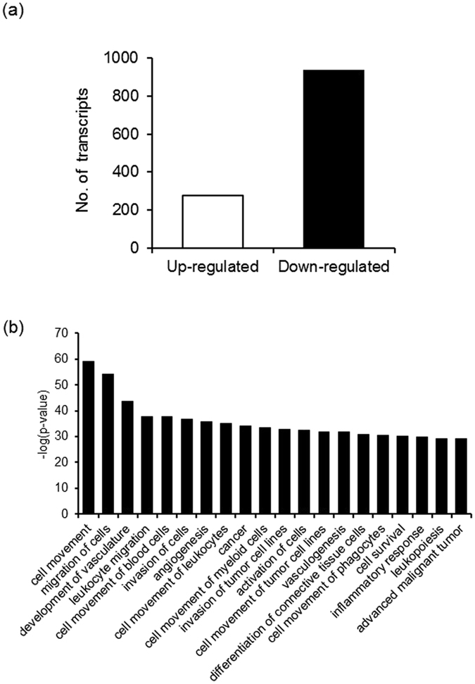
During antral folliculogenesis, developmental competence of prospective oocytes is regulated in large part by the follicular somatic component to prepare the oocyte for the final stage of maturation and subsequent embryo development. The underlying molecular mechanisms are poorly understood. Oocytes reaching the advanced stage of follicular growth by administration of exogenous follicle-stimulating hormone (FSH) possess higher developmental competence than oocytes in FSH-untreated smaller follicles. In this study, the transcriptomic profile of the cumulus cells from cows receiving FSH administration (FSH-priming) was compared, as a model of high oocyte competence, with that from untreated donor cows (control). Ingenuity Pathway Analysis showed that cumulus cells receiving FSH-priming were rich in down-regulated transcripts associated with cell movement and migration, including the extracellular matrix-related transcripts, probably preventing the disruption of cell-to-cell contacts. Interestingly, the transcriptomic profile of up-regulated genes in the control group was similar to that of granulosa cells from atretic follicles. Interferon regulatory factor 7 was activated as the key upstream regulator of FSH-priming. Thus, acquisition of developmental competence by oocytes can be ensured by the integrity of cumulus cells involved in cell-to-cell communication and cell survival, which may help achieve enhanced oocyte-somatic cell coupling.


Conflict of interest statement

The authors declare that they have no competing interests.

Figures

Figure 1
Figure 1
Extracellular expansion of the cumulus cells and developmental competence of oocytes from both unpriming and FSH priming cows. The cumulus expansion index of the COCs derived from donor cows without (control) or with FSH-priming, which were cultured without the ligand (none) or with amphiregulin (AREG), was examined at 22 h of oocyte maturation (a). Effect of FSH-priming prior to OPU and supplementation of FSH during in vitro maturation on oocyte developmental competence were examined by measuring the following on-time embryo development milestones; cleaved embryos at day 2 (b) and blastocyst development on day 7 (c).
Figure 2
Figure 2
FSH-priming-sensitive transcripts and biological function in bovine cumulus cells. Number of FSH-priming-sensitive transcripts were defined as (FSH-priming_FRKM + 0.1)/(Control_FRKM + 0.1) ≥ 2 and FSH-priming_FRKM ≥ 1 (up-regulated) and (FSH-priming_FRKM + 0.1)/(Control_FRKM + 0.1) ≤ 0.5 and Control_FRKM ≥ 1 (down-regulated) (a). FSH-priming-sensitive biological functions were identified by Ingenuity Pathway Analysis (IPA). The top 20 functions for which an activation z score was ≤ −2.0 and −log(p-value) > 1 are shown as decreased biofunctions (b). The functions were identified and ranked by their −log(p-value). The significance of functions was defined as −log(p-value) > 1.
Figure 3
Figure 3
Upstream regulator analysis of FSH-priming-sensitive transcripts in cumulus cells. Ingenuity Pathway Analysis was used for the prediction of upstream molecules including cytokines, growth factors, transcription factors and chemicals. Selected upstream molecules, from the top 30, whose predicted activation status was “inhibited” (a) or “activated” (b) with corresponding molecules are shown. Inhibited and activated upstream molecules were defined as an activation z-score ≤ −1.5 and −log(P-value) > 1, and as an activation z-score ≥ 1.5 and −log(p-value) > 1, respectively. The upstream molecules were ranked by their −log(p-value). The significance was defined as −log(p-value) > 1. Brown, pink, and blue lines indicate upstream regulators, and up-regulated and down-regulated genes stimulated by each upstream molecule, respectively.
Figure 4
Figure 4
Canonical pathway analysis in bovine cumulus cells with FSH-priming. Ingenuity Pathway Analysis was used for predicting canonical pathways. The canonical pathways are identified and ranked by their z-score and -log(P-value). Down-regulated (a) and up-regulated canonical pathways (b) by FSH-priming were defined as a z-score ≤ −1.5 and −log(P-value) > 1, and as a z-score ≥ 1.5 and −log(P-value) > 1, respectively. The significance of canonical pathway was defined as −log(P-value) > 1.
Figure 5
Figure 5
Validation of RNA-seq results by qPCR. Selected down-regulated (a) and up-regulated FSH-priming-sensitive genes (b) in bovine cumulus cells are shown. mRNA expression levels in the cumulus cells derived from eight donor cows without (control) or with FSH-priming (FSH) are represented as a box-and-whisker plots. Boxes reflect two quartiles, the 25th and 75th percentiles, and the interior horizontal line indicates the median. Whiskers indicate the maximum and minimum values within the acceptable range defined by the two quartiles. Open circles denote outliers. Crosses indicate mean value. Asterisks indicate significant difference at P < 0.05. TGFB1, transforming growth factor beta 1; PDGFB, platelet-derived growth factor subunit B; HBEGF, heparin-binding epidermal growth factor-like growth factor; STAT3, signal transducers and activator of transcription 3; TGFBR2, transforming growth factor beta receptor 2; HAS2, hyaluronan synthase 2; TNFAIP6, tumour necrosis factor, alpha-induced protein 6; PTX3, pentraxin 3; THBS1, thrombospondin 1; MMP2, matrix metalloproteinase 2; BMPR1B, bone morphogenetic protein receptor type 1B; IRF7, Interferon regulatory factor; ISG15, interferon-stimulated gene 15; OAS1X, 2′-5′-oligoadenylate synthetase 1, 40/46 kDa; MX1, interferon-induced GTP-binding protein; STAT1, signal transducers and activator of transcription 3; CYP19A1, cytochrome p450 family 19 subfamily a member 1.
Figure 6
Figure 6
Hypothetical signalling network and status in bovine cumulus cells modified by FSH-priming based on RNA-seq expression signature. FSH-priming stimulates an anti-inflammatory cascade via the activation of IRF7 pathway following TGFB1 pathway inhibition. The inhibited TGFB1 pathway may result from the inhibition of MEK signalling. FSH-priming prevents cell movement/migration via the inhibition of genes involved in extracellular matrix expansion (HAS2, TNFAIP6, and PTX3) following MEK signalling inhibition, which may be related to enhancing cell-to-cell communication. Moreover, FSH PRIMING stimulates cAMP-PKA, which may participate in the inhibition of MEK signalling and activation of cell-to-cell communication such as gap-junctional communication (a). Cumulus cells in a low competence model without FSH-priming are in a state of poor cell-to-cell communication, progressing atresia, and driving a spontaneous ovulation-like cascade. Conversely, cumulus cells in a high competence model following FSH-priming are in a state of enhanced cell-to-cell communication and are maintained healthy by the activated anti-inflammatory cascade, which allow the transport of nutritional molecules from cumulus cells to oocytes at the time of final oocyte maturation (b). MEK, mitogen-activated protein kinase/extracellular signal-regulated kinase kinase; cAMP-PKA, cyclic adenosine monophosphate-protein kinase A signalling pathway.
Figure 7
Figure 7
Schematic representation of experimental design. The first OPU from small to medium follicles was performed on unstimulated animals on arbitrary days of the oestrus cycle (control). Then, follicles larger than 8 mm in diameter were ablated (DF-A), and CIDRs were inserted on day 5 (the day of first OPU = day 0). The cows then received 30 mg FSH twice a day from days 7 to 10, in decreasing doses (6, 6, 4, 4, 3, 3, 3, and 2 mg) by intramuscular injection. Cloprostenol (PGF2α; 0.75 mg) was administered in the morning of day 9. The second OPU was performed 48 h after PGF2α administration (day 11), and only follicles larger than 5 mm in diameter (medium and large) were aspirated. CIDRs were removed from cows just before OPU (FSH-priming). COCs of only morphological category A and B were used for investigating cumulus expansion and developmental competence of oocytes and analysing the RNA-seq and real-time qPCR of cumulus cells.

Similar articles

Cited by

References

    1. Gilchrist RB, Thompson JG. Oocyte maturation: emerging concepts and technologies to improve developmental potential in vitro. Theriogenology. 2007;67:6–15. doi: 10.1016/j.theriogenology.2006.09.027. - DOI - PubMed
    1. De Vos M, Smitz J, Thompson JG, Gilchrist RB. The definition of IVM is clear-variations need defining. Human reproduction (Oxford, England) 2016;31:2411–2415. doi: 10.1093/humrep/dew208. - DOI - PubMed
    1. Erickson GF, Wang C, Hsueh AJ. FSH induction of functional LH receptors in granulosa cells cultured in a chemically defined medium. Nature. 1979;279:336–338. doi: 10.1038/279336a0. - DOI - PubMed
    1. El-Hayek S, Demeestere I, Clarke HJ. Follicle-stimulating hormone regulates expression and activity of epidermal growth factor receptor in the murine ovarian follicle. Proceedings of the National Academy of Sciences of the United States of America. 2014;111:16778–16783. doi: 10.1073/pnas.1414648111. - DOI - PMC - PubMed
    1. El-Hayek S, Clarke HJ. Follicle-Stimulating Hormone Increases Gap Junctional Communication Between Somatic and Germ-Line Follicular Compartments During Murine Oogenesis. Biology of reproduction. 2015;93:47. doi: 10.1095/biolreprod.115.129569. - DOI - PubMed

Publication types

LinkOut - more resources